Magnesium »
PDB 4cgl-4cs4 »
4cok »
Magnesium in PDB 4cok: Functional and Structural Characterization of Pyruvate Decarboxylase From Gluconoacetobacter Diazotrophicus
Enzymatic activity of Functional and Structural Characterization of Pyruvate Decarboxylase From Gluconoacetobacter Diazotrophicus
All present enzymatic activity of Functional and Structural Characterization of Pyruvate Decarboxylase From Gluconoacetobacter Diazotrophicus:
4.1.1.1;
4.1.1.1;
Protein crystallography data
The structure of Functional and Structural Characterization of Pyruvate Decarboxylase From Gluconoacetobacter Diazotrophicus, PDB code: 4cok
was solved by
L.J.Vanzyl,
W.-D.Schubert,
M.Tuffin,
D.A.Cowan,
with X-Ray Crystallography technique. A brief refinement statistics is given in the table below:
| Resolution Low / High (Å) | 84.06 / 1.69 |
| Space group | C 1 2 1 |
| Cell size a, b, c (Å), α, β, γ (°) | 129.070, 140.960, 91.060, 90.00, 125.78, 90.00 |
| R / Rfree (%) | 13.19 / 16.294 |
Magnesium Binding Sites:
The binding sites of Magnesium atom in the Functional and Structural Characterization of Pyruvate Decarboxylase From Gluconoacetobacter Diazotrophicus
(pdb code 4cok). This binding sites where shown within
5.0 Angstroms radius around Magnesium atom.
In total 2 binding sites of Magnesium where determined in the Functional and Structural Characterization of Pyruvate Decarboxylase From Gluconoacetobacter Diazotrophicus, PDB code: 4cok:
Jump to Magnesium binding site number: 1; 2;
In total 2 binding sites of Magnesium where determined in the Functional and Structural Characterization of Pyruvate Decarboxylase From Gluconoacetobacter Diazotrophicus, PDB code: 4cok:
Jump to Magnesium binding site number: 1; 2;
Magnesium binding site 1 out of 2 in 4cok
Go back to
Magnesium binding site 1 out
of 2 in the Functional and Structural Characterization of Pyruvate Decarboxylase From Gluconoacetobacter Diazotrophicus
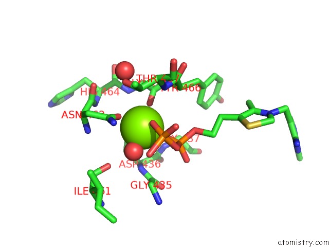
Mono view
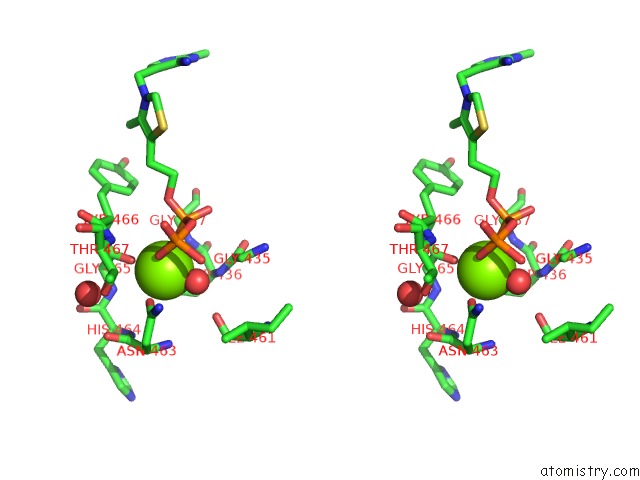
Stereo pair view
Mono view
Stereo pair view
A full contact list of Magnesium with other atoms in the Mg binding
site number 1 of Functional and Structural Characterization of Pyruvate Decarboxylase From Gluconoacetobacter Diazotrophicus within 5.0Å range:
|
Magnesium binding site 2 out of 2 in 4cok
Go back to
Magnesium binding site 2 out
of 2 in the Functional and Structural Characterization of Pyruvate Decarboxylase From Gluconoacetobacter Diazotrophicus
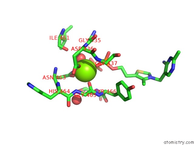
Mono view
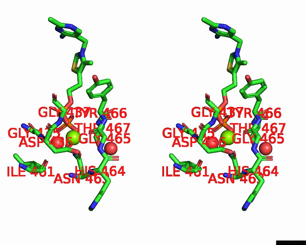
Stereo pair view
Mono view
Stereo pair view
A full contact list of Magnesium with other atoms in the Mg binding
site number 2 of Functional and Structural Characterization of Pyruvate Decarboxylase From Gluconoacetobacter Diazotrophicus within 5.0Å range:
|
Reference:
L.J.Vanzyl,
W.-D.Schubert,
M.Tuffin,
D.A.Cowan.
Functional and Structural Characterization of Pyruvate Decarboxylase From Gluconoacetobacter Diazotrophicus To Be Published.
Page generated: Mon Aug 11 07:17:35 2025
Last articles
Mg in 4JHDMg in 4JH6
Mg in 4JH8
Mg in 4JH7
Mg in 4JH3
Mg in 4JH5
Mg in 4JF2
Mg in 4JH2
Mg in 4JH1
Mg in 4JEJ