Magnesium »
PDB 4ct4-4d4i »
4d05 »
Magnesium in PDB 4d05: Structure and Activity of A Minimal-Type Atp-Dependent Dna Ligase From A Psychrotolernt Bacterium
Protein crystallography data
The structure of Structure and Activity of A Minimal-Type Atp-Dependent Dna Ligase From A Psychrotolernt Bacterium, PDB code: 4d05
was solved by
A.Williamson,
U.Rothweiler,
H.-K.S.Leiros,
with X-Ray Crystallography technique. A brief refinement statistics is given in the table below:
| Resolution Low / High (Å) | 24.610 / 1.65 |
| Space group | C 1 2 1 |
| Cell size a, b, c (Å), α, β, γ (°) | 178.427, 43.976, 89.633, 90.00, 105.92, 90.00 |
| R / Rfree (%) | 14.54 / 19.24 |
Magnesium Binding Sites:
The binding sites of Magnesium atom in the Structure and Activity of A Minimal-Type Atp-Dependent Dna Ligase From A Psychrotolernt Bacterium
(pdb code 4d05). This binding sites where shown within
5.0 Angstroms radius around Magnesium atom.
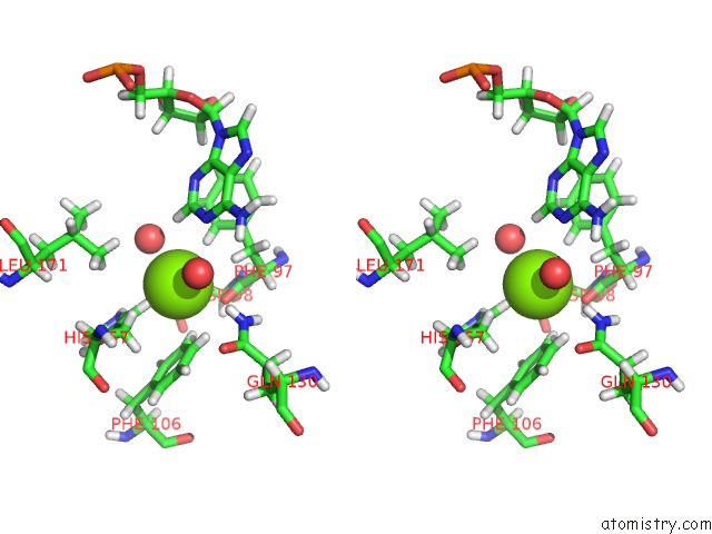
In total only one binding site of Magnesium was determined in the Structure and Activity of A Minimal-Type Atp-Dependent Dna Ligase From A Psychrotolernt Bacterium, PDB code: 4d05:
In total only one binding site of Magnesium was determined in the Structure and Activity of A Minimal-Type Atp-Dependent Dna Ligase From A Psychrotolernt Bacterium, PDB code: 4d05:
Magnesium binding site 1 out of 1 in 4d05
Go back to
Magnesium binding site 1 out
of 1 in the Structure and Activity of A Minimal-Type Atp-Dependent Dna Ligase From A Psychrotolernt Bacterium
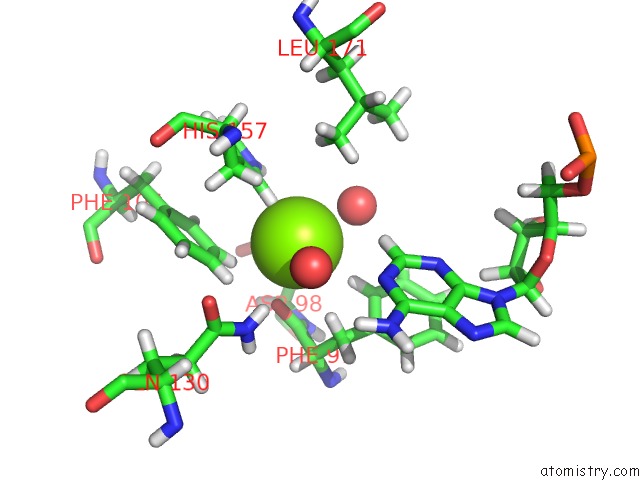
Mono view
Stereo pair view
Mono view
Stereo pair view
A full contact list of Magnesium with other atoms in the Mg binding
site number 1 of Structure and Activity of A Minimal-Type Atp-Dependent Dna Ligase From A Psychrotolernt Bacterium within 5.0Å range:
|
Reference:
A.Williamson,
U.Rothweiler,
H.-K.S.Leiros.
Enzyme-Adenylate Structure of A Bacterial Atp-Dependent Dna Ligase with A Minimized Dna-Binding Surface Acta Crystallogr.,Sect.D V. 70 3043 2014.
ISSN: ISSN 0907-4449
DOI: 10.1107/S1399004714021099
Page generated: Mon Aug 11 07:22:29 2025
ISSN: ISSN 0907-4449
DOI: 10.1107/S1399004714021099
Last articles
Mg in 4ZEXMg in 4ZEW
Mg in 4ZEV
Mg in 4ZES
Mg in 4ZCW
Mg in 4ZDQ
Mg in 4ZDK
Mg in 4ZD6
Mg in 4ZDJ
Mg in 4ZCM