Magnesium »
PDB 4d5e-4dhf »
4dck »
Magnesium in PDB 4dck: Crystal Structure of the C-Terminus of Voltage-Gated Sodium Channel in Complex with FGF13 and Cam
Protein crystallography data
The structure of Crystal Structure of the C-Terminus of Voltage-Gated Sodium Channel in Complex with FGF13 and Cam, PDB code: 4dck
was solved by
B.C.Chung,
C.Wang,
H.Yan,
G.S.Pitt,
S.Y.Lee,
with X-Ray Crystallography technique. A brief refinement statistics is given in the table below:
| Resolution Low / High (Å) | 39.86 / 2.20 |
| Space group | P 21 3 |
| Cell size a, b, c (Å), α, β, γ (°) | 126.041, 126.041, 126.041, 90.00, 90.00, 90.00 |
| R / Rfree (%) | 21 / 22.7 |
Magnesium Binding Sites:
The binding sites of Magnesium atom in the Crystal Structure of the C-Terminus of Voltage-Gated Sodium Channel in Complex with FGF13 and Cam
(pdb code 4dck). This binding sites where shown within
5.0 Angstroms radius around Magnesium atom.
In total 3 binding sites of Magnesium where determined in the Crystal Structure of the C-Terminus of Voltage-Gated Sodium Channel in Complex with FGF13 and Cam, PDB code: 4dck:
Jump to Magnesium binding site number: 1; 2; 3;
In total 3 binding sites of Magnesium where determined in the Crystal Structure of the C-Terminus of Voltage-Gated Sodium Channel in Complex with FGF13 and Cam, PDB code: 4dck:
Jump to Magnesium binding site number: 1; 2; 3;
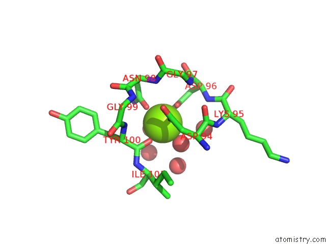
Magnesium binding site 1 out of 3 in 4dck
Go back to
Magnesium binding site 1 out
of 3 in the Crystal Structure of the C-Terminus of Voltage-Gated Sodium Channel in Complex with FGF13 and Cam
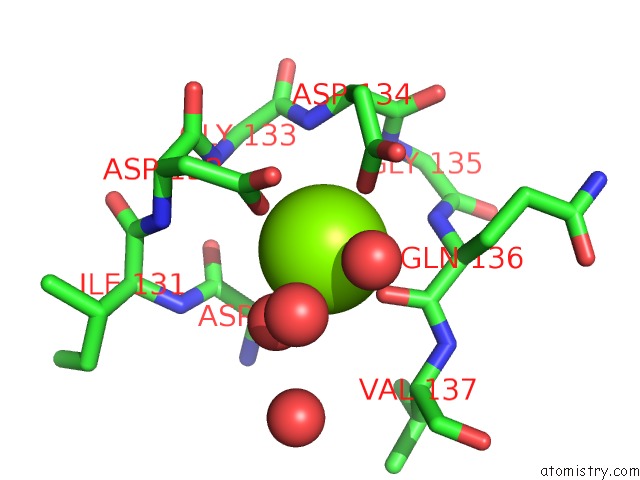
Mono view
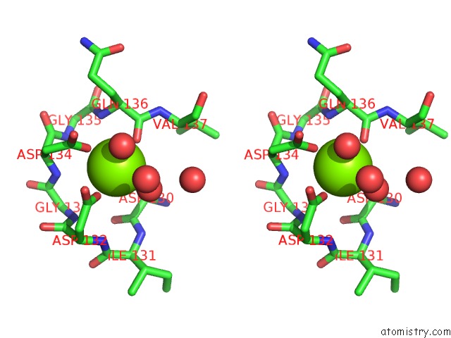
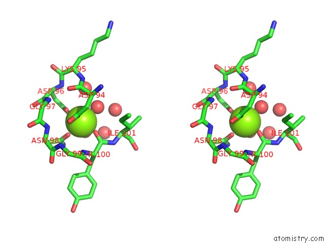
Stereo pair view
Mono view
Stereo pair view
A full contact list of Magnesium with other atoms in the Mg binding
site number 1 of Crystal Structure of the C-Terminus of Voltage-Gated Sodium Channel in Complex with FGF13 and Cam within 5.0Å range:
|
Magnesium binding site 2 out of 3 in 4dck
Go back to
Magnesium binding site 2 out
of 3 in the Crystal Structure of the C-Terminus of Voltage-Gated Sodium Channel in Complex with FGF13 and Cam
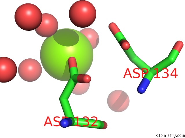
Mono view
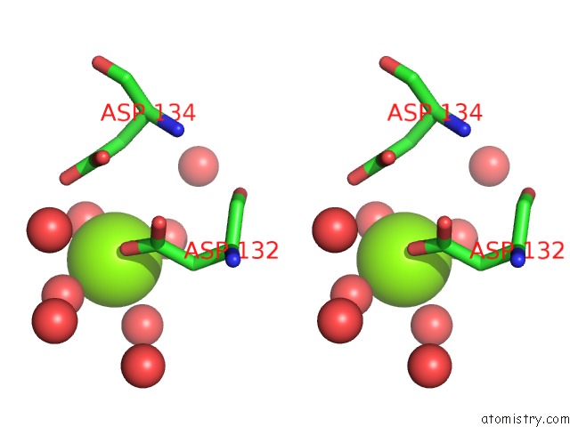
Stereo pair view
Mono view
Stereo pair view
A full contact list of Magnesium with other atoms in the Mg binding
site number 2 of Crystal Structure of the C-Terminus of Voltage-Gated Sodium Channel in Complex with FGF13 and Cam within 5.0Å range:
|
Magnesium binding site 3 out of 3 in 4dck
Go back to
Magnesium binding site 3 out
of 3 in the Crystal Structure of the C-Terminus of Voltage-Gated Sodium Channel in Complex with FGF13 and Cam
Mono view
Stereo pair view
Mono view
Stereo pair view
A full contact list of Magnesium with other atoms in the Mg binding
site number 3 of Crystal Structure of the C-Terminus of Voltage-Gated Sodium Channel in Complex with FGF13 and Cam within 5.0Å range:
|
Reference:
C.Wang,
B.C.Chung,
H.Yan,
S.Y.Lee,
G.S.Pitt.
Crystal Structure of the Ternary Complex of A Nav C-Terminal Domain, A Fibroblast Growth Factor Homologous Factor, and Calmodulin. Structure V. 20 1167 2012.
ISSN: ISSN 0969-2126
PubMed: 22705208
DOI: 10.1016/J.STR.2012.05.001
Page generated: Mon Aug 11 07:26:58 2025
ISSN: ISSN 0969-2126
PubMed: 22705208
DOI: 10.1016/J.STR.2012.05.001
Last articles
Mg in 4FI4Mg in 4FIG
Mg in 4FI1
Mg in 4FI3
Mg in 4FHX
Mg in 4FHY
Mg in 4FHV
Mg in 4FHW
Mg in 4FFR
Mg in 4FH5