Magnesium »
PDB 4doa-4dv5 »
4dr2 »
Magnesium in PDB 4dr2: Crystal Structure of the Thermus Thermophilus (HB8) 30S Ribosomal Subunit with Multiple Copies of Paromomycin Molecules Bound
Protein crystallography data
The structure of Crystal Structure of the Thermus Thermophilus (HB8) 30S Ribosomal Subunit with Multiple Copies of Paromomycin Molecules Bound, PDB code: 4dr2
was solved by
H.Demirci,
F.Murphy Iv,
E.Murphy,
S.T.Gregory,
A.E.Dahlberg,
G.Jogl,
with X-Ray Crystallography technique. A brief refinement statistics is given in the table below:
| Resolution Low / High (Å) | 44.39 / 3.25 |
| Space group | P 41 21 2 |
| Cell size a, b, c (Å), α, β, γ (°) | 402.596, 402.596, 177.577, 90.00, 90.00, 90.00 |
| R / Rfree (%) | 17.1 / 21.1 |
Other elements in 4dr2:
The structure of Crystal Structure of the Thermus Thermophilus (HB8) 30S Ribosomal Subunit with Multiple Copies of Paromomycin Molecules Bound also contains other interesting chemical elements:
| Zinc | (Zn) | 2 atoms |
Magnesium Binding Sites:
Pages:
>>> Page 1 <<< Page 2, Binding sites: 11 - 20; Page 3, Binding sites: 21 - 30; Page 4, Binding sites: 31 - 40; Page 5, Binding sites: 41 - 50; Page 6, Binding sites: 51 - 60; Page 7, Binding sites: 61 - 70; Page 8, Binding sites: 71 - 80; Page 9, Binding sites: 81 - 90; Page 10, Binding sites: 91 - 100; Page 11, Binding sites: 101 - 110; Page 12, Binding sites: 111 - 120; Page 13, Binding sites: 121 - 130; Page 14, Binding sites: 131 - 140; Page 15, Binding sites: 141 - 150; Page 16, Binding sites: 151 - 160; Page 17, Binding sites: 161 - 170; Page 18, Binding sites: 171 - 180; Page 19, Binding sites: 181 - 190; Page 20, Binding sites: 191 - 200; Page 21, Binding sites: 201 - 210; Page 22, Binding sites: 211 - 220; Page 23, Binding sites: 221 - 230; Page 24, Binding sites: 231 - 240; Page 25, Binding sites: 241 - 250; Page 26, Binding sites: 251 - 260; Page 27, Binding sites: 261 - 270; Page 28, Binding sites: 271 - 280; Page 29, Binding sites: 281 - 290; Page 30, Binding sites: 291 - 300; Page 31, Binding sites: 301 - 310; Page 32, Binding sites: 311 - 320; Page 33, Binding sites: 321 - 327;Binding sites:
The binding sites of Magnesium atom in the Crystal Structure of the Thermus Thermophilus (HB8) 30S Ribosomal Subunit with Multiple Copies of Paromomycin Molecules Bound (pdb code 4dr2). This binding sites where shown within 5.0 Angstroms radius around Magnesium atom.In total 327 binding sites of Magnesium where determined in the Crystal Structure of the Thermus Thermophilus (HB8) 30S Ribosomal Subunit with Multiple Copies of Paromomycin Molecules Bound, PDB code: 4dr2:
Jump to Magnesium binding site number: 1; 2; 3; 4; 5; 6; 7; 8; 9; 10;
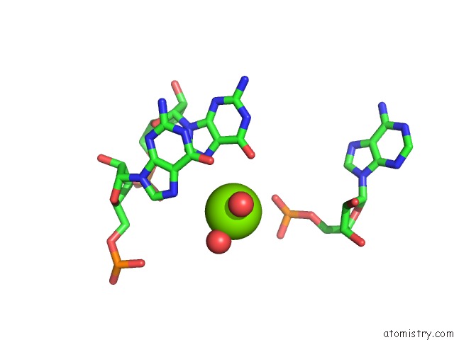
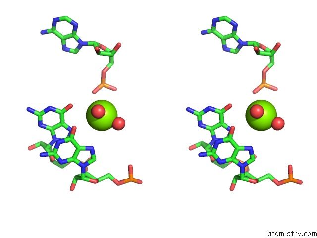
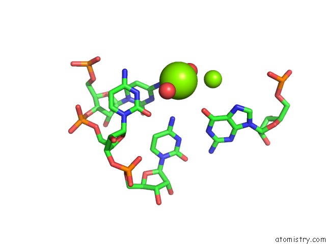
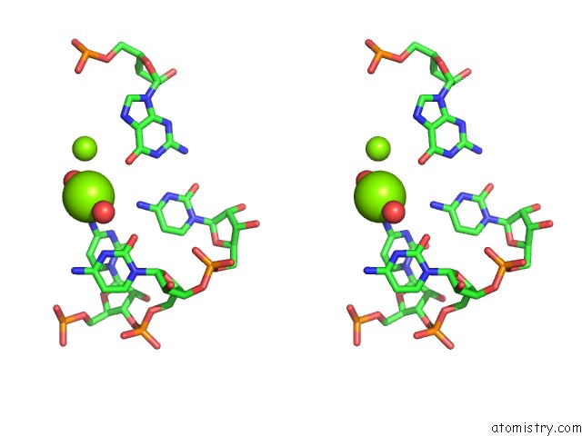
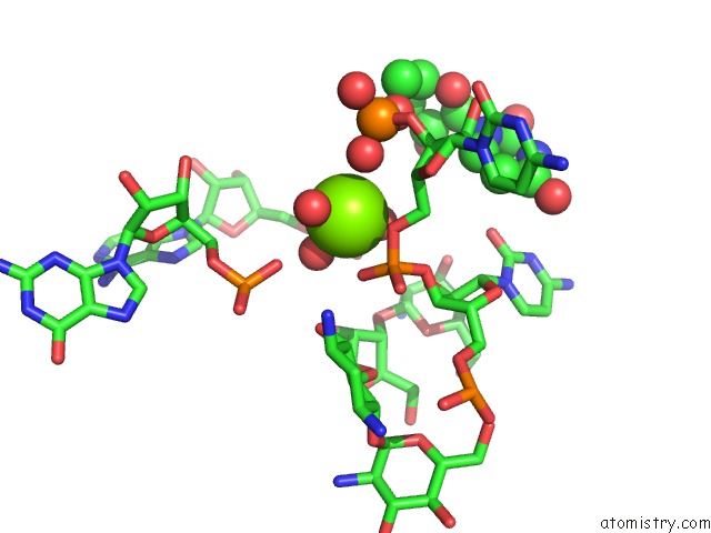
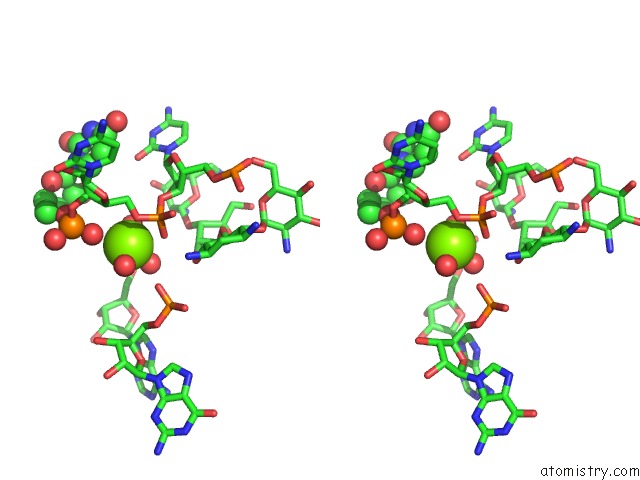
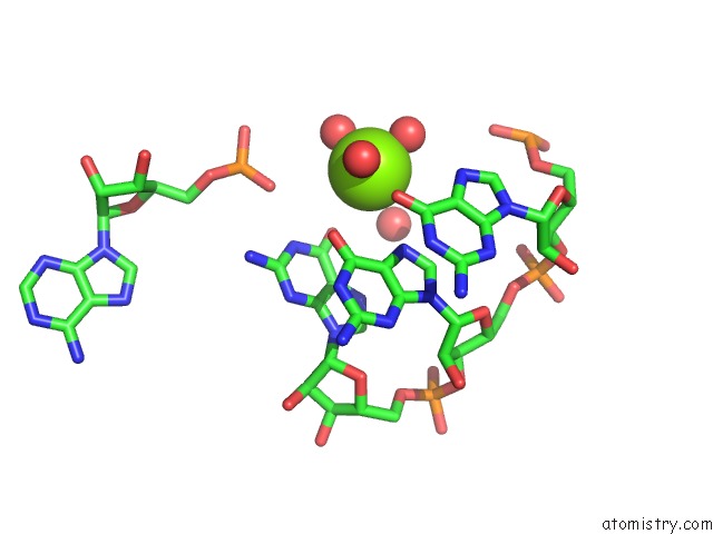
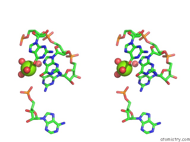
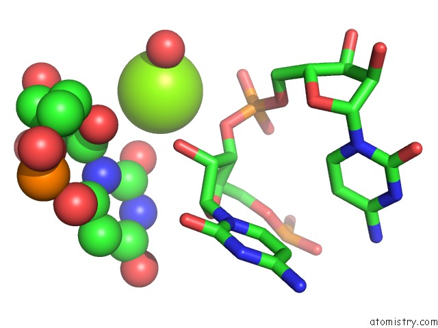
Magnesium binding site 1 out of 327 in 4dr2
Go back to
Magnesium binding site 1 out
of 327 in the Crystal Structure of the Thermus Thermophilus (HB8) 30S Ribosomal Subunit with Multiple Copies of Paromomycin Molecules Bound
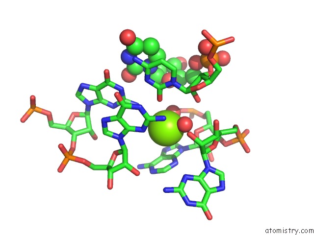
Mono view
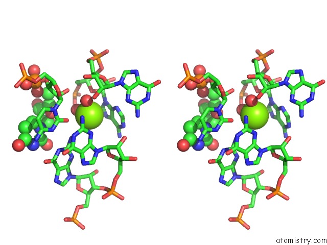
Stereo pair view
Mono view
Stereo pair view
A full contact list of Magnesium with other atoms in the Mg binding
site number 1 of Crystal Structure of the Thermus Thermophilus (HB8) 30S Ribosomal Subunit with Multiple Copies of Paromomycin Molecules Bound within 5.0Å range:
|
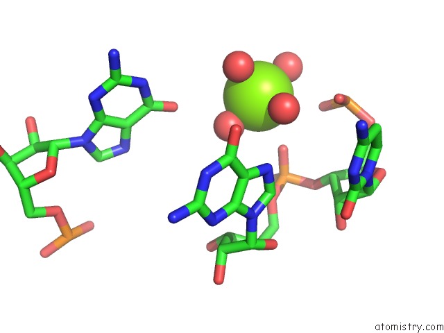
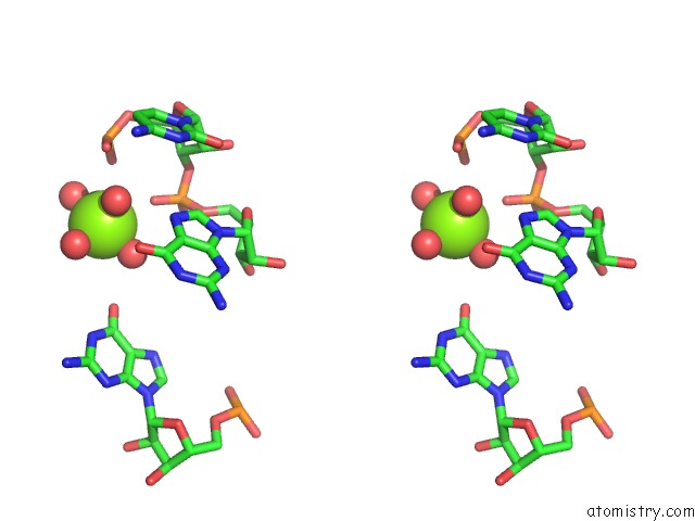
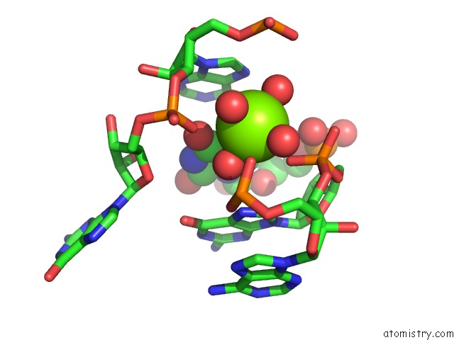
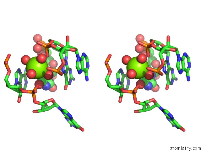
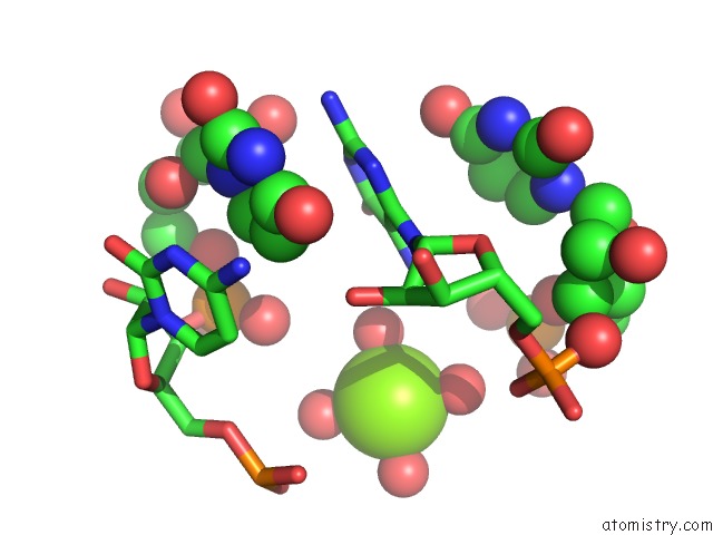
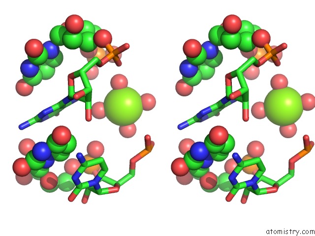
Magnesium binding site 2 out of 327 in 4dr2
Go back to
Magnesium binding site 2 out
of 327 in the Crystal Structure of the Thermus Thermophilus (HB8) 30S Ribosomal Subunit with Multiple Copies of Paromomycin Molecules Bound
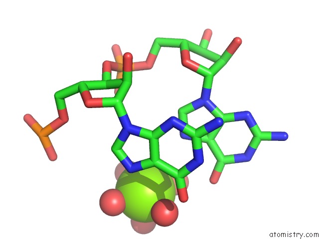
Mono view
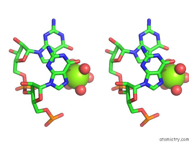
Stereo pair view
Mono view
Stereo pair view
A full contact list of Magnesium with other atoms in the Mg binding
site number 2 of Crystal Structure of the Thermus Thermophilus (HB8) 30S Ribosomal Subunit with Multiple Copies of Paromomycin Molecules Bound within 5.0Å range:
|
Magnesium binding site 3 out of 327 in 4dr2
Go back to
Magnesium binding site 3 out
of 327 in the Crystal Structure of the Thermus Thermophilus (HB8) 30S Ribosomal Subunit with Multiple Copies of Paromomycin Molecules Bound
Mono view
Stereo pair view
Mono view
Stereo pair view
A full contact list of Magnesium with other atoms in the Mg binding
site number 3 of Crystal Structure of the Thermus Thermophilus (HB8) 30S Ribosomal Subunit with Multiple Copies of Paromomycin Molecules Bound within 5.0Å range:
|
Magnesium binding site 4 out of 327 in 4dr2
Go back to
Magnesium binding site 4 out
of 327 in the Crystal Structure of the Thermus Thermophilus (HB8) 30S Ribosomal Subunit with Multiple Copies of Paromomycin Molecules Bound
Mono view
Stereo pair view
Mono view
Stereo pair view
A full contact list of Magnesium with other atoms in the Mg binding
site number 4 of Crystal Structure of the Thermus Thermophilus (HB8) 30S Ribosomal Subunit with Multiple Copies of Paromomycin Molecules Bound within 5.0Å range:
|
Magnesium binding site 5 out of 327 in 4dr2
Go back to
Magnesium binding site 5 out
of 327 in the Crystal Structure of the Thermus Thermophilus (HB8) 30S Ribosomal Subunit with Multiple Copies of Paromomycin Molecules Bound
Mono view
Stereo pair view
Mono view
Stereo pair view
A full contact list of Magnesium with other atoms in the Mg binding
site number 5 of Crystal Structure of the Thermus Thermophilus (HB8) 30S Ribosomal Subunit with Multiple Copies of Paromomycin Molecules Bound within 5.0Å range:
|
Magnesium binding site 6 out of 327 in 4dr2
Go back to
Magnesium binding site 6 out
of 327 in the Crystal Structure of the Thermus Thermophilus (HB8) 30S Ribosomal Subunit with Multiple Copies of Paromomycin Molecules Bound
Mono view
Stereo pair view
Mono view
Stereo pair view
A full contact list of Magnesium with other atoms in the Mg binding
site number 6 of Crystal Structure of the Thermus Thermophilus (HB8) 30S Ribosomal Subunit with Multiple Copies of Paromomycin Molecules Bound within 5.0Å range:
|
Magnesium binding site 7 out of 327 in 4dr2
Go back to
Magnesium binding site 7 out
of 327 in the Crystal Structure of the Thermus Thermophilus (HB8) 30S Ribosomal Subunit with Multiple Copies of Paromomycin Molecules Bound
Mono view
Stereo pair view
Mono view
Stereo pair view
A full contact list of Magnesium with other atoms in the Mg binding
site number 7 of Crystal Structure of the Thermus Thermophilus (HB8) 30S Ribosomal Subunit with Multiple Copies of Paromomycin Molecules Bound within 5.0Å range:
|
Magnesium binding site 8 out of 327 in 4dr2
Go back to
Magnesium binding site 8 out
of 327 in the Crystal Structure of the Thermus Thermophilus (HB8) 30S Ribosomal Subunit with Multiple Copies of Paromomycin Molecules Bound
Mono view
Stereo pair view
Mono view
Stereo pair view
A full contact list of Magnesium with other atoms in the Mg binding
site number 8 of Crystal Structure of the Thermus Thermophilus (HB8) 30S Ribosomal Subunit with Multiple Copies of Paromomycin Molecules Bound within 5.0Å range:
|
Magnesium binding site 9 out of 327 in 4dr2
Go back to
Magnesium binding site 9 out
of 327 in the Crystal Structure of the Thermus Thermophilus (HB8) 30S Ribosomal Subunit with Multiple Copies of Paromomycin Molecules Bound
Mono view
Stereo pair view
Mono view
Stereo pair view
A full contact list of Magnesium with other atoms in the Mg binding
site number 9 of Crystal Structure of the Thermus Thermophilus (HB8) 30S Ribosomal Subunit with Multiple Copies of Paromomycin Molecules Bound within 5.0Å range:
|
Magnesium binding site 10 out of 327 in 4dr2
Go back to
Magnesium binding site 10 out
of 327 in the Crystal Structure of the Thermus Thermophilus (HB8) 30S Ribosomal Subunit with Multiple Copies of Paromomycin Molecules Bound
Mono view

Stereo pair view
Mono view
Stereo pair view
A full contact list of Magnesium with other atoms in the Mg binding
site number 10 of Crystal Structure of the Thermus Thermophilus (HB8) 30S Ribosomal Subunit with Multiple Copies of Paromomycin Molecules Bound within 5.0Å range:
|
Reference:
H.Demirci,
F.Murphy,
E.Murphy,
S.T.Gregory,
A.E.Dahlberg,
G.Jogl.
A Structural Basis For Streptomycin-Induced Misreading of the Genetic Code. Nat Commun V. 4 1355 2013.
ISSN: ESSN 2041-1723
PubMed: 23322043
DOI: 10.1038/NCOMMS2346
Page generated: Thu Aug 15 17:20:52 2024
ISSN: ESSN 2041-1723
PubMed: 23322043
DOI: 10.1038/NCOMMS2346
Last articles
Zn in 9J0NZn in 9J0O
Zn in 9J0P
Zn in 9FJX
Zn in 9EKB
Zn in 9C0F
Zn in 9CAH
Zn in 9CH0
Zn in 9CH3
Zn in 9CH1