Magnesium »
PDB 4dv7-4e7z »
4e4f »
Magnesium in PDB 4e4f: Crystal Structure of Enolase PC1_0802 (Target Efi-502240) From Pectobacterium Carotovorum Subsp. Carotovorum PC1
Protein crystallography data
The structure of Crystal Structure of Enolase PC1_0802 (Target Efi-502240) From Pectobacterium Carotovorum Subsp. Carotovorum PC1, PDB code: 4e4f
was solved by
Y.Patskovsky,
R.Toro,
R.Bhosle,
B.Hillerich,
R.D.Seidel,
E.Washington,
A.Scott Glenn,
S.Chowdhury,
B.Evans,
J.Hammonds,
W.D.Zencheck,
H.J.Imker,
J.A.Gerlt,
S.C.Almo,
Enzyme Function Initiative (Efi),
with X-Ray Crystallography technique. A brief refinement statistics is given in the table below:
| Resolution Low / High (Å) | 50.00 / 2.00 |
| Space group | C 1 2 1 |
| Cell size a, b, c (Å), α, β, γ (°) | 204.278, 86.338, 114.463, 90.00, 123.07, 90.00 |
| R / Rfree (%) | 18.8 / 24.5 |
Other elements in 4e4f:
The structure of Crystal Structure of Enolase PC1_0802 (Target Efi-502240) From Pectobacterium Carotovorum Subsp. Carotovorum PC1 also contains other interesting chemical elements:
| Chlorine | (Cl) | 2 atoms |
Magnesium Binding Sites:
The binding sites of Magnesium atom in the Crystal Structure of Enolase PC1_0802 (Target Efi-502240) From Pectobacterium Carotovorum Subsp. Carotovorum PC1
(pdb code 4e4f). This binding sites where shown within
5.0 Angstroms radius around Magnesium atom.
In total 4 binding sites of Magnesium where determined in the Crystal Structure of Enolase PC1_0802 (Target Efi-502240) From Pectobacterium Carotovorum Subsp. Carotovorum PC1, PDB code: 4e4f:
Jump to Magnesium binding site number: 1; 2; 3; 4;
In total 4 binding sites of Magnesium where determined in the Crystal Structure of Enolase PC1_0802 (Target Efi-502240) From Pectobacterium Carotovorum Subsp. Carotovorum PC1, PDB code: 4e4f:
Jump to Magnesium binding site number: 1; 2; 3; 4;
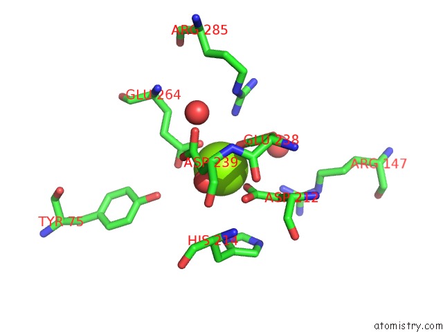
Magnesium binding site 1 out of 4 in 4e4f
Go back to
Magnesium binding site 1 out
of 4 in the Crystal Structure of Enolase PC1_0802 (Target Efi-502240) From Pectobacterium Carotovorum Subsp. Carotovorum PC1
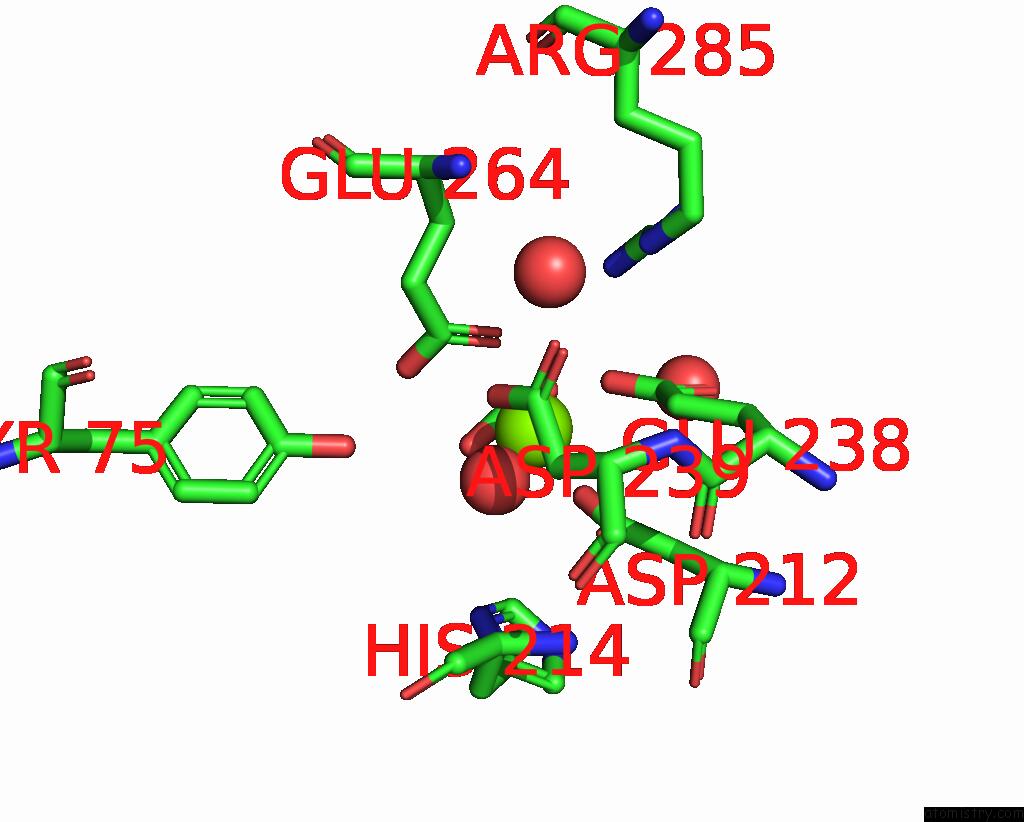
Mono view
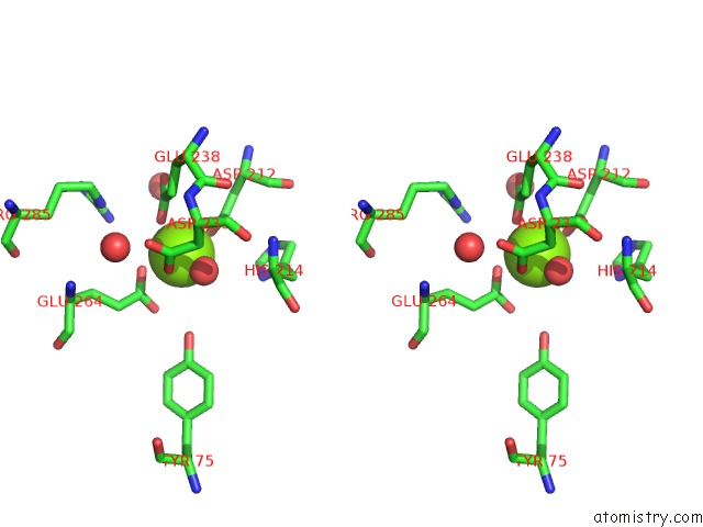
Stereo pair view
Mono view
Stereo pair view
A full contact list of Magnesium with other atoms in the Mg binding
site number 1 of Crystal Structure of Enolase PC1_0802 (Target Efi-502240) From Pectobacterium Carotovorum Subsp. Carotovorum PC1 within 5.0Å range:
|
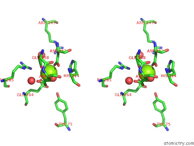
Magnesium binding site 2 out of 4 in 4e4f
Go back to
Magnesium binding site 2 out
of 4 in the Crystal Structure of Enolase PC1_0802 (Target Efi-502240) From Pectobacterium Carotovorum Subsp. Carotovorum PC1
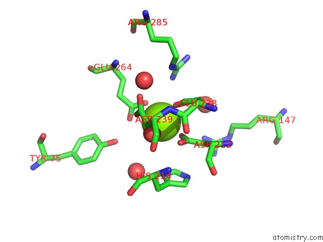
Mono view
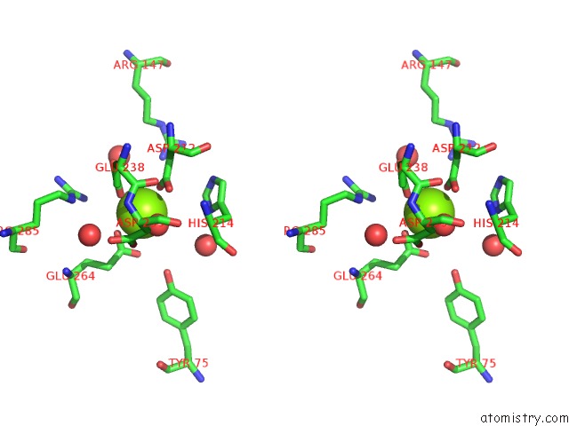
Stereo pair view
Mono view
Stereo pair view
A full contact list of Magnesium with other atoms in the Mg binding
site number 2 of Crystal Structure of Enolase PC1_0802 (Target Efi-502240) From Pectobacterium Carotovorum Subsp. Carotovorum PC1 within 5.0Å range:
|
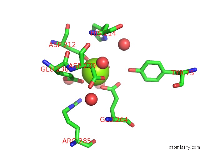
Magnesium binding site 3 out of 4 in 4e4f
Go back to
Magnesium binding site 3 out
of 4 in the Crystal Structure of Enolase PC1_0802 (Target Efi-502240) From Pectobacterium Carotovorum Subsp. Carotovorum PC1
Mono view
Stereo pair view
Mono view
Stereo pair view
A full contact list of Magnesium with other atoms in the Mg binding
site number 3 of Crystal Structure of Enolase PC1_0802 (Target Efi-502240) From Pectobacterium Carotovorum Subsp. Carotovorum PC1 within 5.0Å range:
|
Magnesium binding site 4 out of 4 in 4e4f
Go back to
Magnesium binding site 4 out
of 4 in the Crystal Structure of Enolase PC1_0802 (Target Efi-502240) From Pectobacterium Carotovorum Subsp. Carotovorum PC1
Mono view
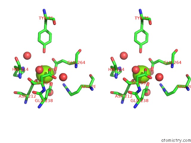
Stereo pair view
Mono view
Stereo pair view
A full contact list of Magnesium with other atoms in the Mg binding
site number 4 of Crystal Structure of Enolase PC1_0802 (Target Efi-502240) From Pectobacterium Carotovorum Subsp. Carotovorum PC1 within 5.0Å range:
|
Reference:
Y.Patskovsky,
R.Toro,
R.Bhosle,
B.Hillerich,
R.D.Seidel,
E.Washington,
A.Scott Glenn,
S.Chowdhury,
B.Evans,
J.Hammonds,
W.D.Zencheck,
H.J.Imker,
J.A.Gerlt,
S.C.Almo.
Crystal Structure of Enolase PC1_0802 From Pectobacterium Carotovorum To Be Published.
Page generated: Mon Aug 11 11:55:58 2025
Last articles
Mg in 4JHDMg in 4JH6
Mg in 4JH8
Mg in 4JH7
Mg in 4JH3
Mg in 4JH5
Mg in 4JF2
Mg in 4JH2
Mg in 4JH1
Mg in 4JEJ