Magnesium »
PDB 4gnr-4gyq »
4gvy »
Magnesium in PDB 4gvy: Crystal Structure of Arginine Kinase in Complex with L-Citrulline and Mgadp
Enzymatic activity of Crystal Structure of Arginine Kinase in Complex with L-Citrulline and Mgadp
All present enzymatic activity of Crystal Structure of Arginine Kinase in Complex with L-Citrulline and Mgadp:
2.7.3.3;
2.7.3.3;
Protein crystallography data
The structure of Crystal Structure of Arginine Kinase in Complex with L-Citrulline and Mgadp, PDB code: 4gvy
was solved by
S.A.Clark,
O.Davulcu,
M.S.Chapman,
with X-Ray Crystallography technique. A brief refinement statistics is given in the table below:
| Resolution Low / High (Å) | 17.64 / 2.09 |
| Space group | P 21 21 21 |
| Cell size a, b, c (Å), α, β, γ (°) | 65.393, 70.312, 80.319, 90.00, 90.00, 90.00 |
| R / Rfree (%) | 17.5 / 21.1 |
Magnesium Binding Sites:
The binding sites of Magnesium atom in the Crystal Structure of Arginine Kinase in Complex with L-Citrulline and Mgadp
(pdb code 4gvy). This binding sites where shown within
5.0 Angstroms radius around Magnesium atom.
In total only one binding site of Magnesium was determined in the Crystal Structure of Arginine Kinase in Complex with L-Citrulline and Mgadp, PDB code: 4gvy:
In total only one binding site of Magnesium was determined in the Crystal Structure of Arginine Kinase in Complex with L-Citrulline and Mgadp, PDB code: 4gvy:
Magnesium binding site 1 out of 1 in 4gvy
Go back to
Magnesium binding site 1 out
of 1 in the Crystal Structure of Arginine Kinase in Complex with L-Citrulline and Mgadp
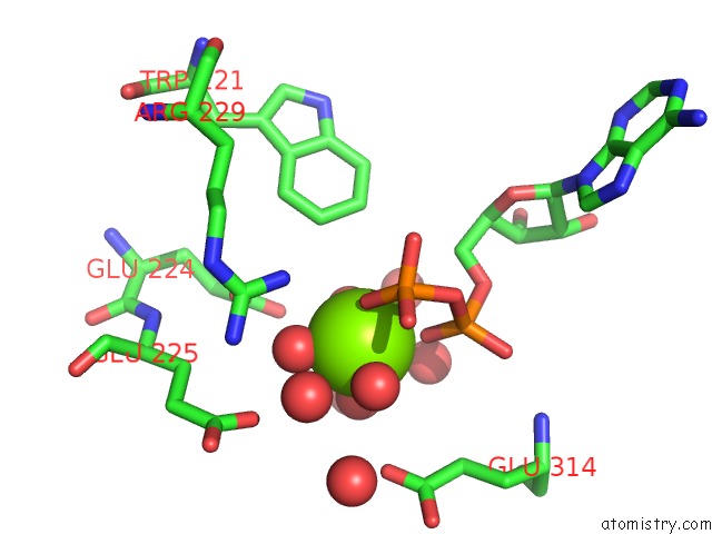
Mono view
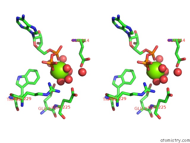
Stereo pair view
Mono view
Stereo pair view
A full contact list of Magnesium with other atoms in the Mg binding
site number 1 of Crystal Structure of Arginine Kinase in Complex with L-Citrulline and Mgadp within 5.0Å range:
|
Reference:
S.A.Clark,
O.Davulcu,
M.S.Chapman.
Crystal Structures of Arginine Kinase in Complex with Adp, Nitrate, and Various Phosphagen Analogs. Biochem.Biophys.Res.Commun. V. 427 212 2012.
ISSN: ISSN 0006-291X
PubMed: 22995310
DOI: 10.1016/J.BBRC.2012.09.053
Page generated: Mon Aug 11 13:38:12 2025
ISSN: ISSN 0006-291X
PubMed: 22995310
DOI: 10.1016/J.BBRC.2012.09.053
Last articles
Mg in 5IWYMg in 5IXT
Mg in 5IWX
Mg in 5IXQ
Mg in 5IXO
Mg in 5IX1
Mg in 5IX2
Mg in 5IWA
Mg in 5IT9
Mg in 5IVG