Magnesium »
PDB 4p9d-4pjj »
4ph5 »
Magnesium in PDB 4ph5: Structure of Human Dna Polymerase Beta Complexed with A Nicked Dna Containing A Ac at N-1 Position and Gc at N Position
Enzymatic activity of Structure of Human Dna Polymerase Beta Complexed with A Nicked Dna Containing A Ac at N-1 Position and Gc at N Position
All present enzymatic activity of Structure of Human Dna Polymerase Beta Complexed with A Nicked Dna Containing A Ac at N-1 Position and Gc at N Position:
2.7.7.7;
2.7.7.7;
Protein crystallography data
The structure of Structure of Human Dna Polymerase Beta Complexed with A Nicked Dna Containing A Ac at N-1 Position and Gc at N Position, PDB code: 4ph5
was solved by
M.C.Koag,
S.Lee,
with X-Ray Crystallography technique. A brief refinement statistics is given in the table below:
| Resolution Low / High (Å) | 20.00 / 2.55 |
| Space group | P 1 21 1 |
| Cell size a, b, c (Å), α, β, γ (°) | 50.613, 80.377, 55.294, 90.00, 107.61, 90.00 |
| R / Rfree (%) | 19.1 / 26.1 |
Other elements in 4ph5:
The structure of Structure of Human Dna Polymerase Beta Complexed with A Nicked Dna Containing A Ac at N-1 Position and Gc at N Position also contains other interesting chemical elements:
| Sodium | (Na) | 2 atoms |
Magnesium Binding Sites:
The binding sites of Magnesium atom in the Structure of Human Dna Polymerase Beta Complexed with A Nicked Dna Containing A Ac at N-1 Position and Gc at N Position
(pdb code 4ph5). This binding sites where shown within
5.0 Angstroms radius around Magnesium atom.
In total 2 binding sites of Magnesium where determined in the Structure of Human Dna Polymerase Beta Complexed with A Nicked Dna Containing A Ac at N-1 Position and Gc at N Position, PDB code: 4ph5:
Jump to Magnesium binding site number: 1; 2;
In total 2 binding sites of Magnesium where determined in the Structure of Human Dna Polymerase Beta Complexed with A Nicked Dna Containing A Ac at N-1 Position and Gc at N Position, PDB code: 4ph5:
Jump to Magnesium binding site number: 1; 2;
Magnesium binding site 1 out of 2 in 4ph5
Go back to
Magnesium binding site 1 out
of 2 in the Structure of Human Dna Polymerase Beta Complexed with A Nicked Dna Containing A Ac at N-1 Position and Gc at N Position
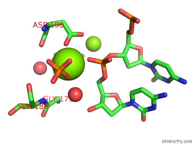
Mono view
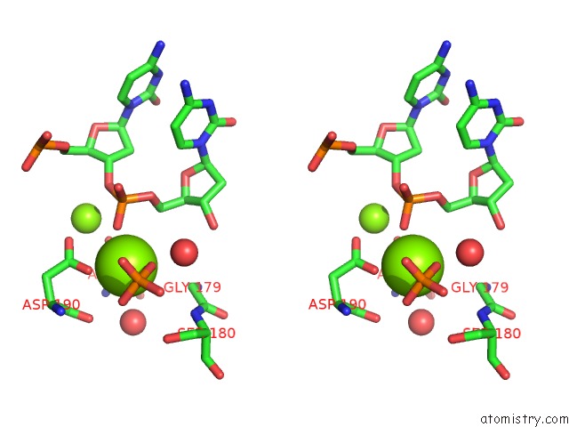
Stereo pair view
Mono view
Stereo pair view
A full contact list of Magnesium with other atoms in the Mg binding
site number 1 of Structure of Human Dna Polymerase Beta Complexed with A Nicked Dna Containing A Ac at N-1 Position and Gc at N Position within 5.0Å range:
|
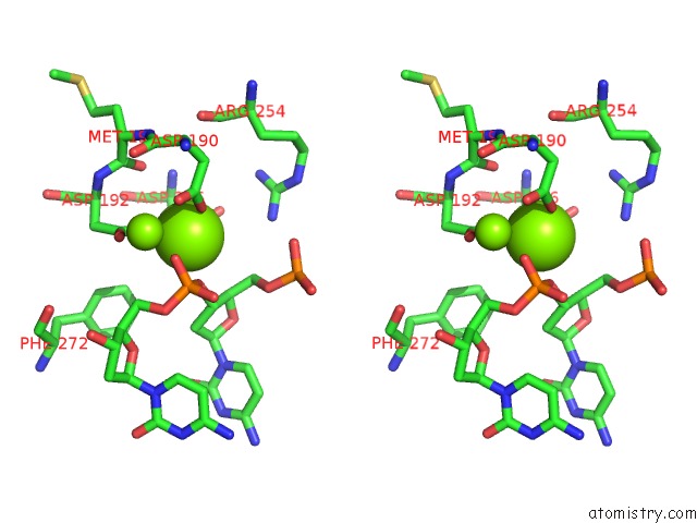
Magnesium binding site 2 out of 2 in 4ph5
Go back to
Magnesium binding site 2 out
of 2 in the Structure of Human Dna Polymerase Beta Complexed with A Nicked Dna Containing A Ac at N-1 Position and Gc at N Position
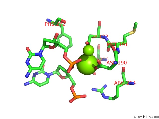
Mono view
Stereo pair view
Mono view
Stereo pair view
A full contact list of Magnesium with other atoms in the Mg binding
site number 2 of Structure of Human Dna Polymerase Beta Complexed with A Nicked Dna Containing A Ac at N-1 Position and Gc at N Position within 5.0Å range:
|
Reference:
M.C.Koag,
S.Lee.
The Spontaneous Base Substitution Mechanisms of Human Dna Polymerase Beta To Be Published.
Page generated: Mon Aug 11 21:54:25 2025
Last articles
Mg in 5JC8Mg in 5JCA
Mg in 5JC7
Mg in 5JC3
Mg in 5JAC
Mg in 5JBH
Mg in 5JBQ
Mg in 5JBG
Mg in 5J9V
Mg in 5JB3