Magnesium »
PDB 4rcz-4rkf »
4ret »
Magnesium in PDB 4ret: Crystal Structure of the Na,K-Atpase E2P-Digoxin Complex with Bound Magnesium
Enzymatic activity of Crystal Structure of the Na,K-Atpase E2P-Digoxin Complex with Bound Magnesium
All present enzymatic activity of Crystal Structure of the Na,K-Atpase E2P-Digoxin Complex with Bound Magnesium:
3.6.3.9;
3.6.3.9;
Protein crystallography data
The structure of Crystal Structure of the Na,K-Atpase E2P-Digoxin Complex with Bound Magnesium, PDB code: 4ret
was solved by
J.L.Gregersen,
M.Laursen,
L.Yatime,
P.Nissen,
N.U.Fedosova,
with X-Ray Crystallography technique. A brief refinement statistics is given in the table below:
| Resolution Low / High (Å) | 49.41 / 4.00 |
| Space group | P 21 21 21 |
| Cell size a, b, c (Å), α, β, γ (°) | 118.240, 118.350, 494.090, 90.00, 90.00, 90.00 |
| R / Rfree (%) | 22.1 / 25.3 |
Magnesium Binding Sites:
The binding sites of Magnesium atom in the Crystal Structure of the Na,K-Atpase E2P-Digoxin Complex with Bound Magnesium
(pdb code 4ret). This binding sites where shown within
5.0 Angstroms radius around Magnesium atom.
In total 6 binding sites of Magnesium where determined in the Crystal Structure of the Na,K-Atpase E2P-Digoxin Complex with Bound Magnesium, PDB code: 4ret:
Jump to Magnesium binding site number: 1; 2; 3; 4; 5; 6;
In total 6 binding sites of Magnesium where determined in the Crystal Structure of the Na,K-Atpase E2P-Digoxin Complex with Bound Magnesium, PDB code: 4ret:
Jump to Magnesium binding site number: 1; 2; 3; 4; 5; 6;
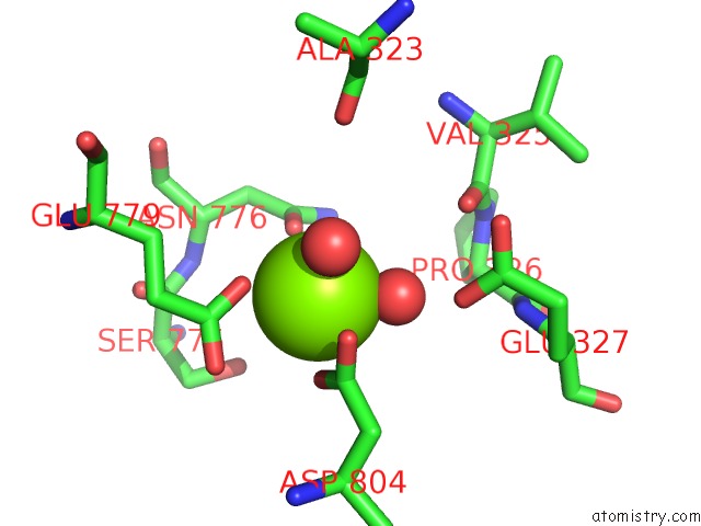
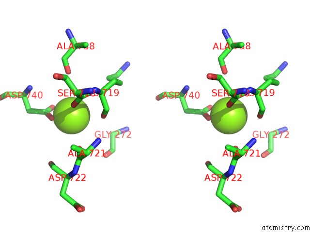
Magnesium binding site 1 out of 6 in 4ret
Go back to
Magnesium binding site 1 out
of 6 in the Crystal Structure of the Na,K-Atpase E2P-Digoxin Complex with Bound Magnesium
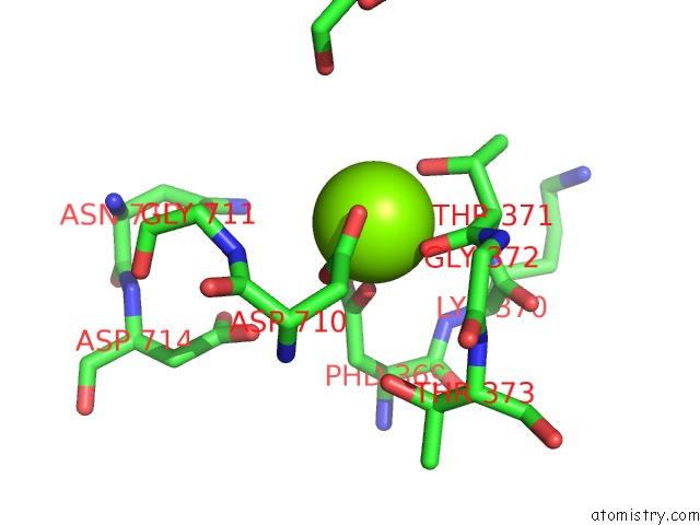
Mono view
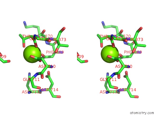
Stereo pair view
Mono view
Stereo pair view
A full contact list of Magnesium with other atoms in the Mg binding
site number 1 of Crystal Structure of the Na,K-Atpase E2P-Digoxin Complex with Bound Magnesium within 5.0Å range:
|
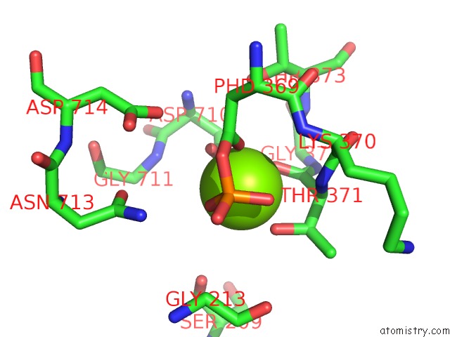
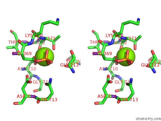
Magnesium binding site 2 out of 6 in 4ret
Go back to
Magnesium binding site 2 out
of 6 in the Crystal Structure of the Na,K-Atpase E2P-Digoxin Complex with Bound Magnesium
Mono view
Stereo pair view
Mono view
Stereo pair view
A full contact list of Magnesium with other atoms in the Mg binding
site number 2 of Crystal Structure of the Na,K-Atpase E2P-Digoxin Complex with Bound Magnesium within 5.0Å range:
|
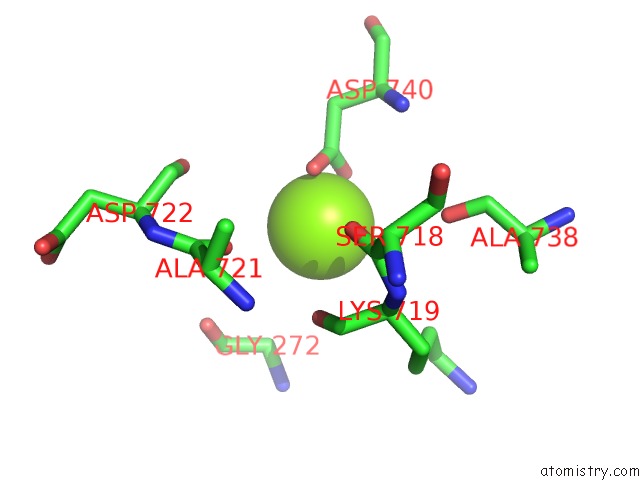
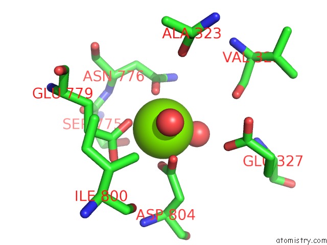
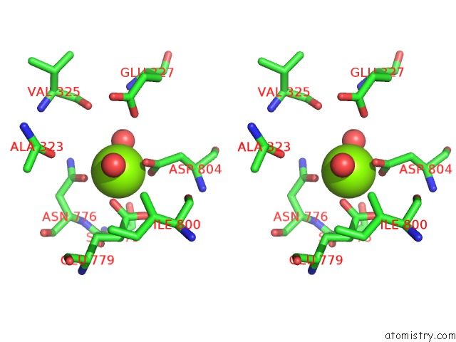
Magnesium binding site 3 out of 6 in 4ret
Go back to
Magnesium binding site 3 out
of 6 in the Crystal Structure of the Na,K-Atpase E2P-Digoxin Complex with Bound Magnesium
Mono view

Stereo pair view
Mono view
Stereo pair view
A full contact list of Magnesium with other atoms in the Mg binding
site number 3 of Crystal Structure of the Na,K-Atpase E2P-Digoxin Complex with Bound Magnesium within 5.0Å range:
|
Magnesium binding site 4 out of 6 in 4ret
Go back to
Magnesium binding site 4 out
of 6 in the Crystal Structure of the Na,K-Atpase E2P-Digoxin Complex with Bound Magnesium
Mono view
Stereo pair view
Mono view
Stereo pair view
A full contact list of Magnesium with other atoms in the Mg binding
site number 4 of Crystal Structure of the Na,K-Atpase E2P-Digoxin Complex with Bound Magnesium within 5.0Å range:
|
Magnesium binding site 5 out of 6 in 4ret
Go back to
Magnesium binding site 5 out
of 6 in the Crystal Structure of the Na,K-Atpase E2P-Digoxin Complex with Bound Magnesium
Mono view
Stereo pair view
Mono view
Stereo pair view
A full contact list of Magnesium with other atoms in the Mg binding
site number 5 of Crystal Structure of the Na,K-Atpase E2P-Digoxin Complex with Bound Magnesium within 5.0Å range:
|
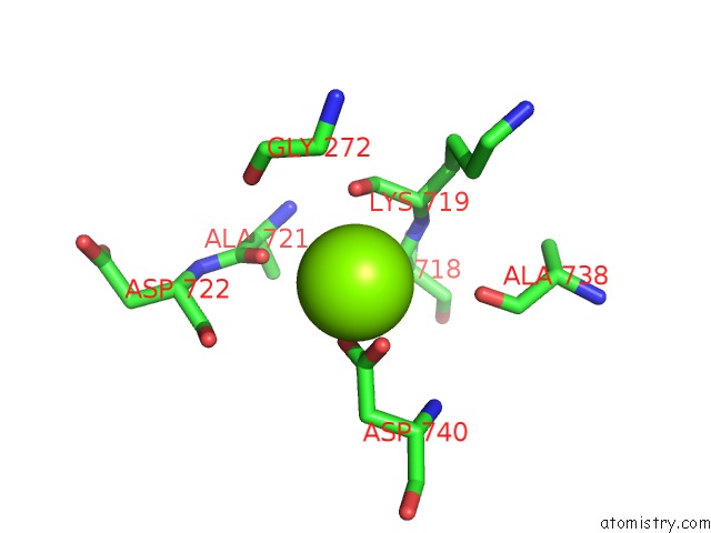
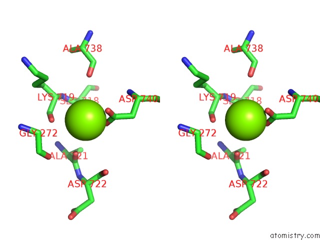
Magnesium binding site 6 out of 6 in 4ret
Go back to
Magnesium binding site 6 out
of 6 in the Crystal Structure of the Na,K-Atpase E2P-Digoxin Complex with Bound Magnesium
Mono view
Stereo pair view
Mono view
Stereo pair view
A full contact list of Magnesium with other atoms in the Mg binding
site number 6 of Crystal Structure of the Na,K-Atpase E2P-Digoxin Complex with Bound Magnesium within 5.0Å range:
|
Reference:
M.Laursen,
J.L.Gregersen,
L.Yatime,
P.Nissen,
N.U.Fedosova.
Structures and Characterization of Digoxin- and Bufalin-Bound Na+,K+-Atpase Compared with the Ouabain-Bound Complex. Proc.Natl.Acad.Sci.Usa 2015.
ISSN: ESSN 1091-6490
PubMed: 25624492
DOI: 10.1073/PNAS.1422997112
Page generated: Mon Aug 11 23:16:50 2025
ISSN: ESSN 1091-6490
PubMed: 25624492
DOI: 10.1073/PNAS.1422997112
Last articles
Mg in 4XSZMg in 4XTJ
Mg in 4XRU
Mg in 4XSY
Mg in 4XSX
Mg in 4XRP
Mg in 4XSH
Mg in 4XSG
Mg in 4XQT
Mg in 4XRH