Magnesium »
PDB 4w5q-4wfn »
4w6d »
Magnesium in PDB 4w6d: Crystal Structure of Full-Length Split Gfp Mutant K26C Disulfide Dimer, P 32 2 1 Space Group, Form 1
Protein crystallography data
The structure of Crystal Structure of Full-Length Split Gfp Mutant K26C Disulfide Dimer, P 32 2 1 Space Group, Form 1, PDB code: 4w6d
was solved by
D.J.Leibly,
G.S.Waldo,
T.O.Yeates,
with X-Ray Crystallography technique. A brief refinement statistics is given in the table below:
| Resolution Low / High (Å) | 87.16 / 3.45 |
| Space group | P 32 2 1 |
| Cell size a, b, c (Å), α, β, γ (°) | 123.110, 123.110, 151.320, 90.00, 90.00, 120.00 |
| R / Rfree (%) | 23.6 / 26.7 |
Magnesium Binding Sites:
The binding sites of Magnesium atom in the Crystal Structure of Full-Length Split Gfp Mutant K26C Disulfide Dimer, P 32 2 1 Space Group, Form 1
(pdb code 4w6d). This binding sites where shown within
5.0 Angstroms radius around Magnesium atom.
In total only one binding site of Magnesium was determined in the Crystal Structure of Full-Length Split Gfp Mutant K26C Disulfide Dimer, P 32 2 1 Space Group, Form 1, PDB code: 4w6d:
In total only one binding site of Magnesium was determined in the Crystal Structure of Full-Length Split Gfp Mutant K26C Disulfide Dimer, P 32 2 1 Space Group, Form 1, PDB code: 4w6d:
Magnesium binding site 1 out of 1 in 4w6d
Go back to
Magnesium binding site 1 out
of 1 in the Crystal Structure of Full-Length Split Gfp Mutant K26C Disulfide Dimer, P 32 2 1 Space Group, Form 1
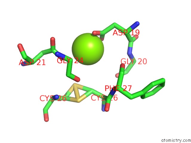
Mono view
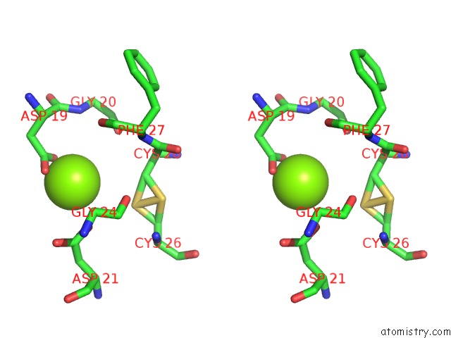
Stereo pair view
Mono view
Stereo pair view
A full contact list of Magnesium with other atoms in the Mg binding
site number 1 of Crystal Structure of Full-Length Split Gfp Mutant K26C Disulfide Dimer, P 32 2 1 Space Group, Form 1 within 5.0Å range:
|
Reference:
D.J.Leibly,
M.A.Arbing,
I.Pashkov,
N.Devore,
T.C.Terwilliger,
T.C.Terwilliger,
T.O.Yeates.
Engineering Novel Oligomeric Gfp Molecules For Synthetic Symmetrization Applications To Be Published.
Page generated: Tue Aug 12 01:10:11 2025
Last articles
Mg in 5CYPMg in 5CYV
Mg in 5CYO
Mg in 5CYR
Mg in 5CX7
Mg in 5CVC
Mg in 5CX6
Mg in 5CUY
Mg in 5CVW
Mg in 5CVH