Magnesium »
PDB 5skf-5t2v »
5svt »
Magnesium in PDB 5svt: Anomalous Cs+ Signal Reveals the Site of Na+ Ion Entry to the Channel Pore of the Human P2X3 Ion Channel Through the Extracellular Fenestrations
Protein crystallography data
The structure of Anomalous Cs+ Signal Reveals the Site of Na+ Ion Entry to the Channel Pore of the Human P2X3 Ion Channel Through the Extracellular Fenestrations, PDB code: 5svt
was solved by
S.E.Mansoor,
W.Lu,
W.Oosterheert,
M.Shekhar,
E.Tajkhorshid,
E.Gouaux,
with X-Ray Crystallography technique. A brief refinement statistics is given in the table below:
| Resolution Low / High (Å) | 47.73 / 3.79 |
| Space group | H 3 2 |
| Cell size a, b, c (Å), α, β, γ (°) | 119.950, 119.950, 236.410, 90.00, 90.00, 120.00 |
| R / Rfree (%) | 27.6 / 31.2 |
Other elements in 5svt:
The structure of Anomalous Cs+ Signal Reveals the Site of Na+ Ion Entry to the Channel Pore of the Human P2X3 Ion Channel Through the Extracellular Fenestrations also contains other interesting chemical elements:
| Sodium | (Na) | 1 atom |
Magnesium Binding Sites:
The binding sites of Magnesium atom in the Anomalous Cs+ Signal Reveals the Site of Na+ Ion Entry to the Channel Pore of the Human P2X3 Ion Channel Through the Extracellular Fenestrations
(pdb code 5svt). This binding sites where shown within
5.0 Angstroms radius around Magnesium atom.
In total only one binding site of Magnesium was determined in the Anomalous Cs+ Signal Reveals the Site of Na+ Ion Entry to the Channel Pore of the Human P2X3 Ion Channel Through the Extracellular Fenestrations, PDB code: 5svt:
In total only one binding site of Magnesium was determined in the Anomalous Cs+ Signal Reveals the Site of Na+ Ion Entry to the Channel Pore of the Human P2X3 Ion Channel Through the Extracellular Fenestrations, PDB code: 5svt:
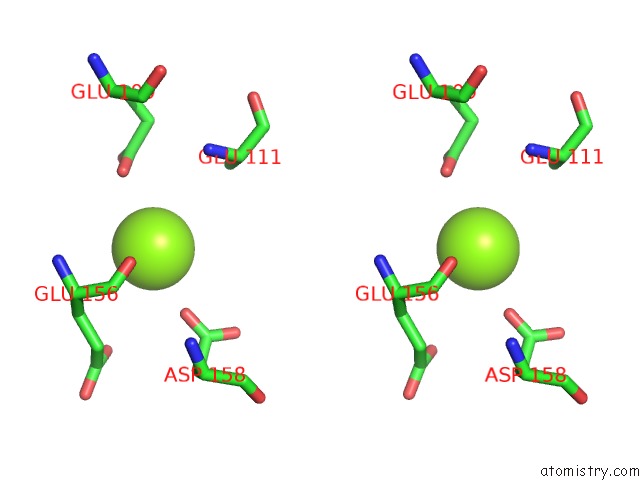
Magnesium binding site 1 out of 1 in 5svt
Go back to
Magnesium binding site 1 out
of 1 in the Anomalous Cs+ Signal Reveals the Site of Na+ Ion Entry to the Channel Pore of the Human P2X3 Ion Channel Through the Extracellular Fenestrations
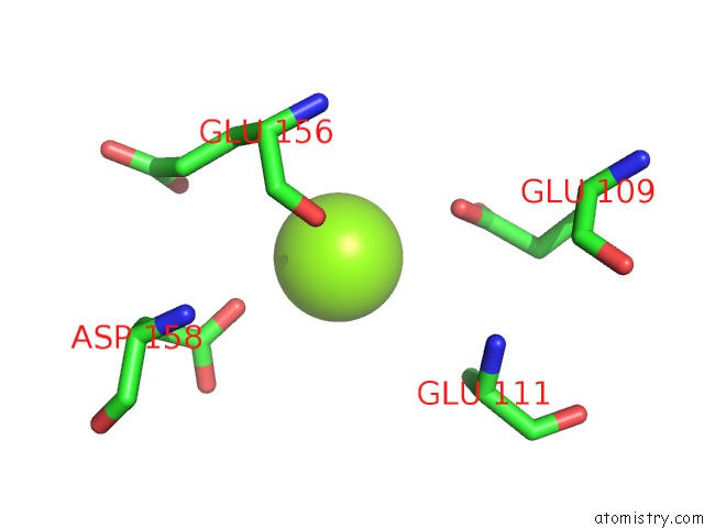
Mono view
Stereo pair view
Mono view
Stereo pair view
A full contact list of Magnesium with other atoms in the Mg binding
site number 1 of Anomalous Cs+ Signal Reveals the Site of Na+ Ion Entry to the Channel Pore of the Human P2X3 Ion Channel Through the Extracellular Fenestrations within 5.0Å range:
|
Reference:
S.E.Mansoor,
W.Lu,
W.Oosterheert,
M.Shekhar,
E.Tajkhorshid,
E.Gouaux.
X-Ray Structures Define Human P2X3 Receptor Gating Cycle and Antagonist Action. Nature V. 538 66 2016.
ISSN: ESSN 1476-4687
PubMed: 27626375
DOI: 10.1038/NATURE19367
Page generated: Mon Sep 30 04:41:04 2024
ISSN: ESSN 1476-4687
PubMed: 27626375
DOI: 10.1038/NATURE19367
Last articles
Zn in 9MJ5Zn in 9HNW
Zn in 9G0L
Zn in 9FNE
Zn in 9DZN
Zn in 9E0I
Zn in 9D32
Zn in 9DAK
Zn in 8ZXC
Zn in 8ZUF