Magnesium »
PDB 6ll8-6ly7 »
6lsn »
Magnesium in PDB 6lsn: Crystal Structure of Tubulin-Inhibitor Complex
Protein crystallography data
The structure of Crystal Structure of Tubulin-Inhibitor Complex, PDB code: 6lsn
was solved by
L.Gang,
Y.X.Wang,
J.J.Cheng,
with X-Ray Crystallography technique. A brief refinement statistics is given in the table below:
| Resolution Low / High (Å) | 33.17 / 2.45 |
| Space group | P 21 21 21 |
| Cell size a, b, c (Å), α, β, γ (°) | 105.296, 158.458, 181.959, 90, 90, 90 |
| R / Rfree (%) | 17.7 / 22 |
Other elements in 6lsn:
The structure of Crystal Structure of Tubulin-Inhibitor Complex also contains other interesting chemical elements:
| Chlorine | (Cl) | 1 atom |
| Calcium | (Ca) | 4 atoms |
Magnesium Binding Sites:
The binding sites of Magnesium atom in the Crystal Structure of Tubulin-Inhibitor Complex
(pdb code 6lsn). This binding sites where shown within
5.0 Angstroms radius around Magnesium atom.
In total 6 binding sites of Magnesium where determined in the Crystal Structure of Tubulin-Inhibitor Complex, PDB code: 6lsn:
Jump to Magnesium binding site number: 1; 2; 3; 4; 5; 6;
In total 6 binding sites of Magnesium where determined in the Crystal Structure of Tubulin-Inhibitor Complex, PDB code: 6lsn:
Jump to Magnesium binding site number: 1; 2; 3; 4; 5; 6;
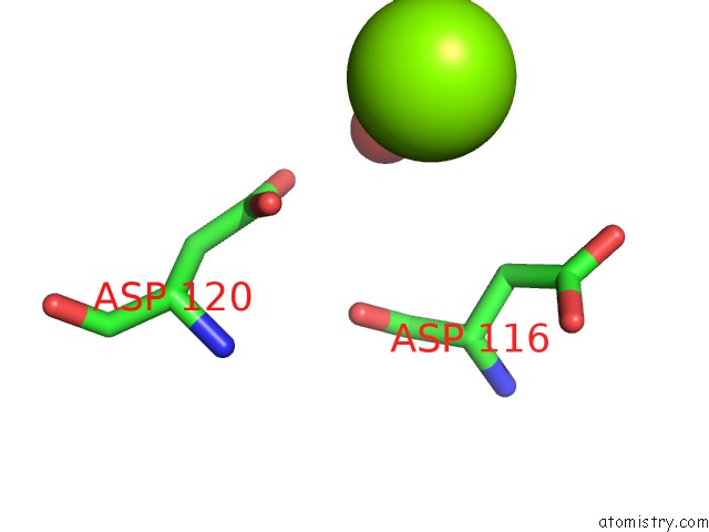
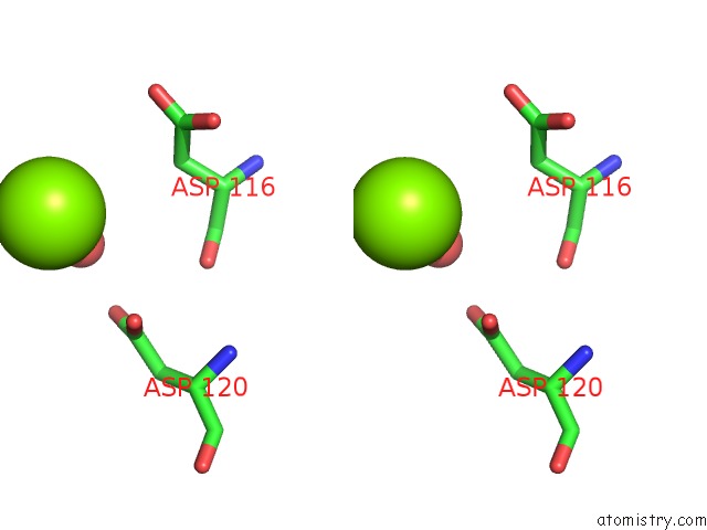
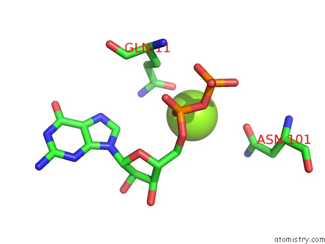
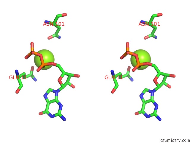
Magnesium binding site 1 out of 6 in 6lsn
Go back to
Magnesium binding site 1 out
of 6 in the Crystal Structure of Tubulin-Inhibitor Complex
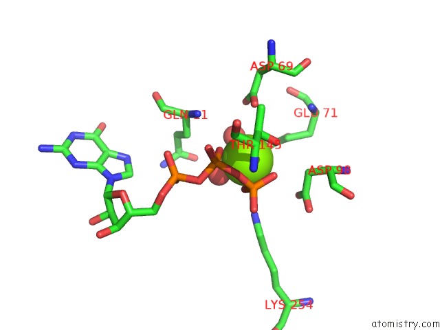
Mono view
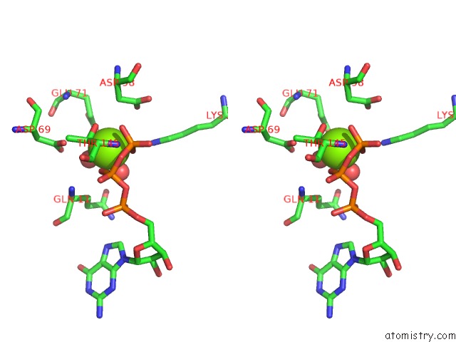
Stereo pair view
Mono view
Stereo pair view
A full contact list of Magnesium with other atoms in the Mg binding
site number 1 of Crystal Structure of Tubulin-Inhibitor Complex within 5.0Å range:
|
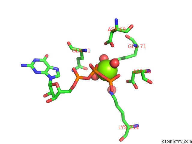
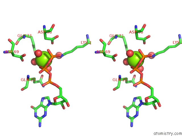
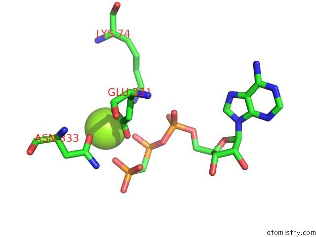
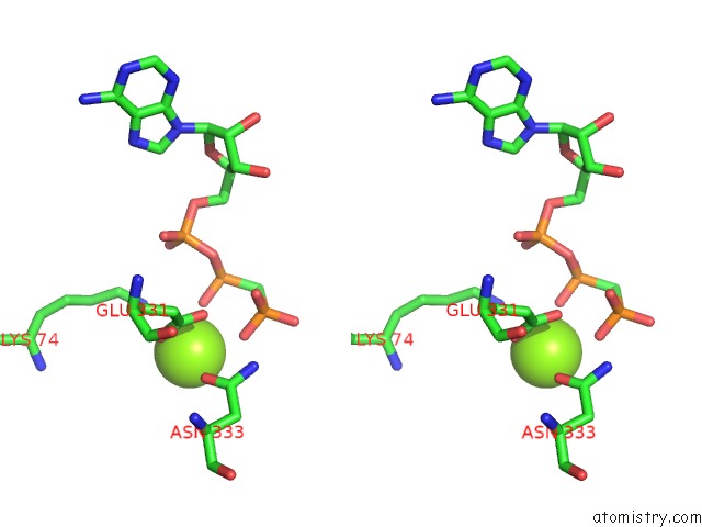
Magnesium binding site 2 out of 6 in 6lsn
Go back to
Magnesium binding site 2 out
of 6 in the Crystal Structure of Tubulin-Inhibitor Complex
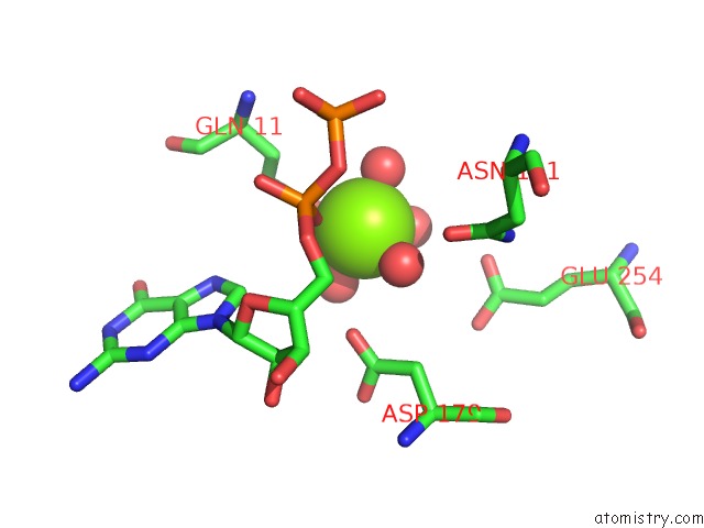
Mono view
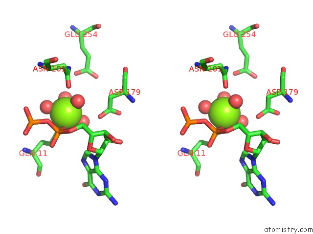
Stereo pair view
Mono view
Stereo pair view
A full contact list of Magnesium with other atoms in the Mg binding
site number 2 of Crystal Structure of Tubulin-Inhibitor Complex within 5.0Å range:
|
Magnesium binding site 3 out of 6 in 6lsn
Go back to
Magnesium binding site 3 out
of 6 in the Crystal Structure of Tubulin-Inhibitor Complex
Mono view
Stereo pair view
Mono view
Stereo pair view
A full contact list of Magnesium with other atoms in the Mg binding
site number 3 of Crystal Structure of Tubulin-Inhibitor Complex within 5.0Å range:
|
Magnesium binding site 4 out of 6 in 6lsn
Go back to
Magnesium binding site 4 out
of 6 in the Crystal Structure of Tubulin-Inhibitor Complex
Mono view
Stereo pair view
Mono view
Stereo pair view
A full contact list of Magnesium with other atoms in the Mg binding
site number 4 of Crystal Structure of Tubulin-Inhibitor Complex within 5.0Å range:
|
Magnesium binding site 5 out of 6 in 6lsn
Go back to
Magnesium binding site 5 out
of 6 in the Crystal Structure of Tubulin-Inhibitor Complex
Mono view
Stereo pair view
Mono view
Stereo pair view
A full contact list of Magnesium with other atoms in the Mg binding
site number 5 of Crystal Structure of Tubulin-Inhibitor Complex within 5.0Å range:
|
Magnesium binding site 6 out of 6 in 6lsn
Go back to
Magnesium binding site 6 out
of 6 in the Crystal Structure of Tubulin-Inhibitor Complex
Mono view
Stereo pair view
Mono view
Stereo pair view
A full contact list of Magnesium with other atoms in the Mg binding
site number 6 of Crystal Structure of Tubulin-Inhibitor Complex within 5.0Å range:
|
Reference:
L.Gang,
Y.X.Wang,
J.J.Chen.
Design, Synthesis, and Bioevaluation of Pyrazolo[1,5-A]Pyrimidine Derivatives As Tubulin Polymerization Inhibitors Targeting the Colchicine Binding Site with Potent Anticancer Activities To Be Published.
Page generated: Wed Aug 13 11:27:37 2025
Last articles
Mg in 7DUJMg in 7DUI
Mg in 7DUH
Mg in 7DUG
Mg in 7DR0
Mg in 7DR1
Mg in 7DU2
Mg in 7DSP
Mg in 7DSJ
Mg in 7DSI