Magnesium »
PDB 7kfr-7klx »
7kjc »
Magnesium in PDB 7kjc: Crystal Structure of the EPHA2 S901E Mutant Intracellular Kd-Sam Domains
Enzymatic activity of Crystal Structure of the EPHA2 S901E Mutant Intracellular Kd-Sam Domains
All present enzymatic activity of Crystal Structure of the EPHA2 S901E Mutant Intracellular Kd-Sam Domains:
2.7.10.1;
2.7.10.1;
Protein crystallography data
The structure of Crystal Structure of the EPHA2 S901E Mutant Intracellular Kd-Sam Domains, PDB code: 7kjc
was solved by
B.C.Lechtenberg,
E.B.Pasquale,
with X-Ray Crystallography technique. A brief refinement statistics is given in the table below:
| Resolution Low / High (Å) | 29.25 / 2.30 |
| Space group | I 1 2 1 |
| Cell size a, b, c (Å), α, β, γ (°) | 121.639, 54.587, 135.538, 90, 95.22, 90 |
| R / Rfree (%) | 18.8 / 22.6 |
Magnesium Binding Sites:
The binding sites of Magnesium atom in the Crystal Structure of the EPHA2 S901E Mutant Intracellular Kd-Sam Domains
(pdb code 7kjc). This binding sites where shown within
5.0 Angstroms radius around Magnesium atom.
In total only one binding site of Magnesium was determined in the Crystal Structure of the EPHA2 S901E Mutant Intracellular Kd-Sam Domains, PDB code: 7kjc:
In total only one binding site of Magnesium was determined in the Crystal Structure of the EPHA2 S901E Mutant Intracellular Kd-Sam Domains, PDB code: 7kjc:
Magnesium binding site 1 out of 1 in 7kjc
Go back to
Magnesium binding site 1 out
of 1 in the Crystal Structure of the EPHA2 S901E Mutant Intracellular Kd-Sam Domains
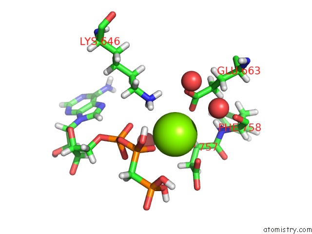
Mono view
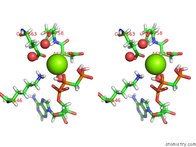
Stereo pair view
Mono view
Stereo pair view
A full contact list of Magnesium with other atoms in the Mg binding
site number 1 of Crystal Structure of the EPHA2 S901E Mutant Intracellular Kd-Sam Domains within 5.0Å range:
|
Reference:
B.C.Lechtenberg,
M.P.Gehring,
E.B.Pasquale.
Crystal Structure of the EPHA2 S901E Mutant Intracellular Kd-Sam Domains To Be Published.
Page generated: Thu Aug 14 08:14:40 2025
Last articles
Mg in 7OUHMg in 7OUG
Mg in 7OUF
Mg in 7OU4
Mg in 7OU0
Mg in 7OTZ
Mg in 7OTP
Mg in 7OTX
Mg in 7OTO
Mg in 7OTJ