Magnesium »
PDB 7x76-7xma »
7xm0 »
Magnesium in PDB 7xm0: Crystal Structure of SAU3AI-C and Dna Substrate Complex
Enzymatic activity of Crystal Structure of SAU3AI-C and Dna Substrate Complex
All present enzymatic activity of Crystal Structure of SAU3AI-C and Dna Substrate Complex:
3.1.21.4;
3.1.21.4;
Protein crystallography data
The structure of Crystal Structure of SAU3AI-C and Dna Substrate Complex, PDB code: 7xm0
was solved by
L.Yahui,
Y.Feng,
H.Jianhua,
with X-Ray Crystallography technique. A brief refinement statistics is given in the table below:
| Resolution Low / High (Å) | 42.23 / 2.60 |
| Space group | P 21 21 21 |
| Cell size a, b, c (Å), α, β, γ (°) | 62.943, 98.391, 209.455, 90, 90, 90 |
| R / Rfree (%) | 18.8 / 23.6 |
Magnesium Binding Sites:
The binding sites of Magnesium atom in the Crystal Structure of SAU3AI-C and Dna Substrate Complex
(pdb code 7xm0). This binding sites where shown within
5.0 Angstroms radius around Magnesium atom.
In total 3 binding sites of Magnesium where determined in the Crystal Structure of SAU3AI-C and Dna Substrate Complex, PDB code: 7xm0:
Jump to Magnesium binding site number: 1; 2; 3;
In total 3 binding sites of Magnesium where determined in the Crystal Structure of SAU3AI-C and Dna Substrate Complex, PDB code: 7xm0:
Jump to Magnesium binding site number: 1; 2; 3;
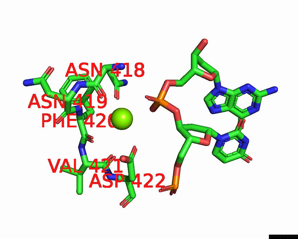
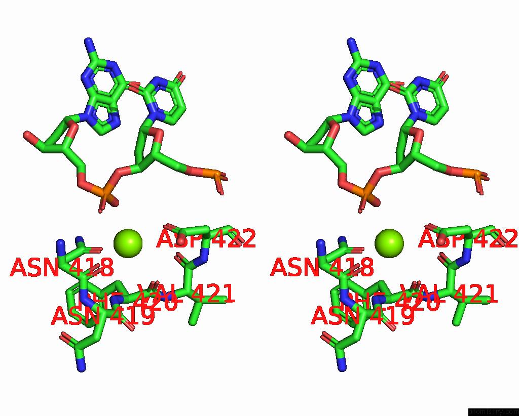
Magnesium binding site 1 out of 3 in 7xm0
Go back to
Magnesium binding site 1 out
of 3 in the Crystal Structure of SAU3AI-C and Dna Substrate Complex
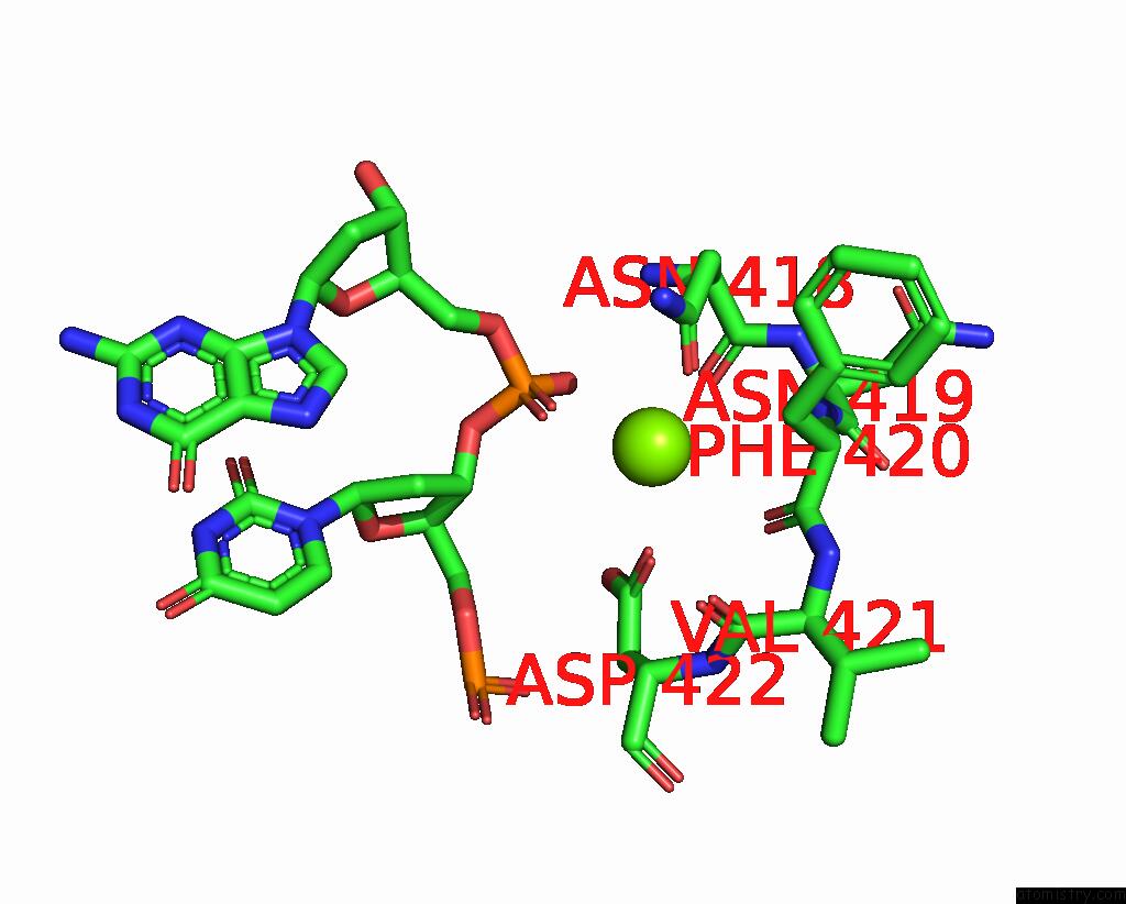
Mono view
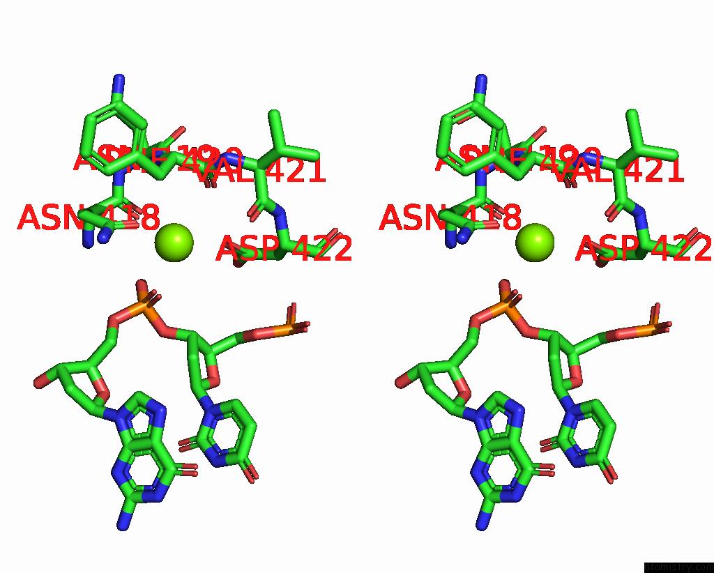
Stereo pair view
Mono view
Stereo pair view
A full contact list of Magnesium with other atoms in the Mg binding
site number 1 of Crystal Structure of SAU3AI-C and Dna Substrate Complex within 5.0Å range:
|
Magnesium binding site 2 out of 3 in 7xm0
Go back to
Magnesium binding site 2 out
of 3 in the Crystal Structure of SAU3AI-C and Dna Substrate Complex
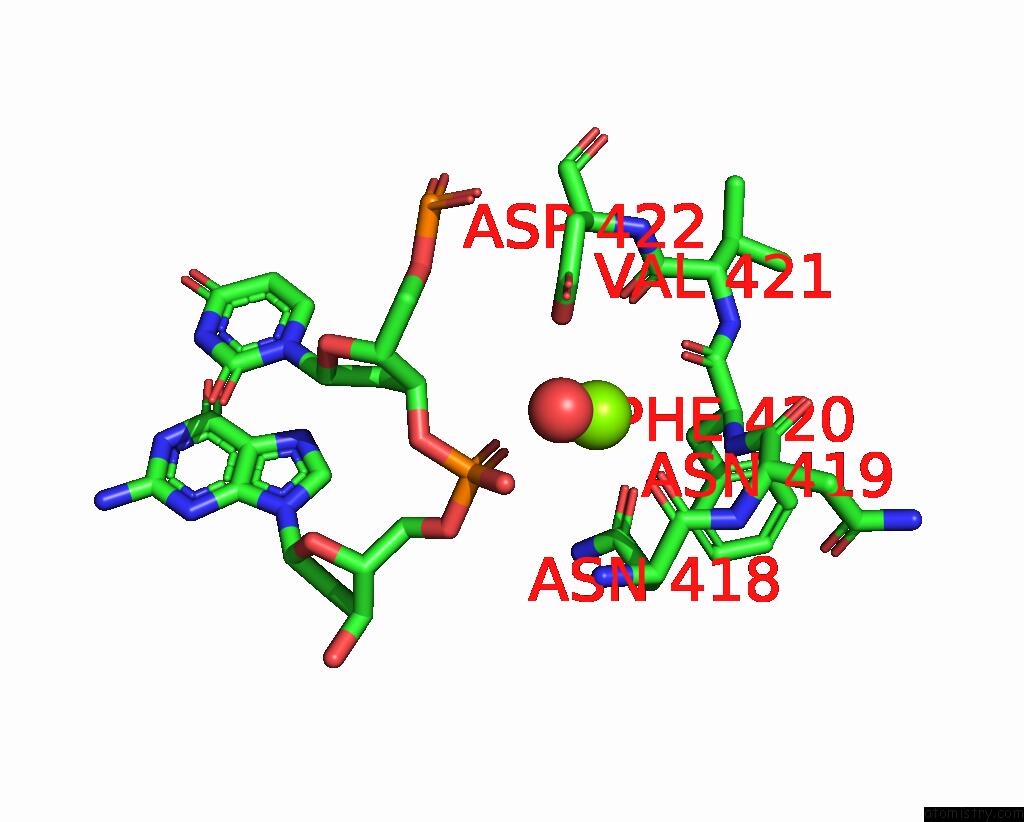
Mono view
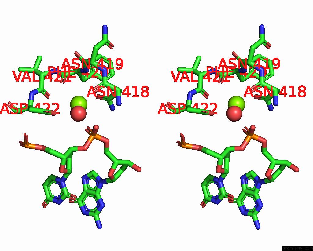
Stereo pair view
Mono view
Stereo pair view
A full contact list of Magnesium with other atoms in the Mg binding
site number 2 of Crystal Structure of SAU3AI-C and Dna Substrate Complex within 5.0Å range:
|
Magnesium binding site 3 out of 3 in 7xm0
Go back to
Magnesium binding site 3 out
of 3 in the Crystal Structure of SAU3AI-C and Dna Substrate Complex
Mono view
Stereo pair view
Mono view
Stereo pair view
A full contact list of Magnesium with other atoms in the Mg binding
site number 3 of Crystal Structure of SAU3AI-C and Dna Substrate Complex within 5.0Å range:
|
Reference:
L.Yahui,
Y.Feng,
H.Jianhua.
The Self-Activation Mechanism of Type Iie Restriction Endonuclease SAU3AI To Be Published.
Page generated: Thu Aug 14 18:28:33 2025
Last articles
Mg in 7Y7BMg in 7Y8A
Mg in 7YGD
Mg in 7YGC
Mg in 7YGB
Mg in 7YGA
Mg in 7YG9
Mg in 7YG8
Mg in 7YG6
Mg in 7YFY