Magnesium »
PDB 8dr6-8e1f »
8dxl »
Magnesium in PDB 8dxl: Hiv-1 Reverse Transcriptase/Rilpivirine with Bound Fragment 4- Iodopyrazole at Multiple Sites
Enzymatic activity of Hiv-1 Reverse Transcriptase/Rilpivirine with Bound Fragment 4- Iodopyrazole at Multiple Sites
All present enzymatic activity of Hiv-1 Reverse Transcriptase/Rilpivirine with Bound Fragment 4- Iodopyrazole at Multiple Sites:
2.7.7.49; 2.7.7.7; 3.1.13.2; 3.1.26.13;
2.7.7.49; 2.7.7.7; 3.1.13.2; 3.1.26.13;
Protein crystallography data
The structure of Hiv-1 Reverse Transcriptase/Rilpivirine with Bound Fragment 4- Iodopyrazole at Multiple Sites, PDB code: 8dxl
was solved by
A.Chopra,
F.X.Ruiz,
J.D.Bauman,
E.Arnold,
with X-Ray Crystallography technique. A brief refinement statistics is given in the table below:
| Resolution Low / High (Å) | 41.20 / 2.25 |
| Space group | C 1 2 1 |
| Cell size a, b, c (Å), α, β, γ (°) | 162.318, 73.314, 109.398, 90, 100.62, 90 |
| R / Rfree (%) | 18.2 / 20.8 |
Other elements in 8dxl:
The structure of Hiv-1 Reverse Transcriptase/Rilpivirine with Bound Fragment 4- Iodopyrazole at Multiple Sites also contains other interesting chemical elements:
| Iodine | (I) | 3 atoms |
Magnesium Binding Sites:
The binding sites of Magnesium atom in the Hiv-1 Reverse Transcriptase/Rilpivirine with Bound Fragment 4- Iodopyrazole at Multiple Sites
(pdb code 8dxl). This binding sites where shown within
5.0 Angstroms radius around Magnesium atom.
In total only one binding site of Magnesium was determined in the Hiv-1 Reverse Transcriptase/Rilpivirine with Bound Fragment 4- Iodopyrazole at Multiple Sites, PDB code: 8dxl:
In total only one binding site of Magnesium was determined in the Hiv-1 Reverse Transcriptase/Rilpivirine with Bound Fragment 4- Iodopyrazole at Multiple Sites, PDB code: 8dxl:
Magnesium binding site 1 out of 1 in 8dxl
Go back to
Magnesium binding site 1 out
of 1 in the Hiv-1 Reverse Transcriptase/Rilpivirine with Bound Fragment 4- Iodopyrazole at Multiple Sites
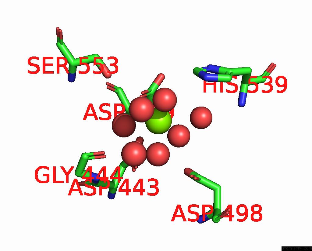
Mono view
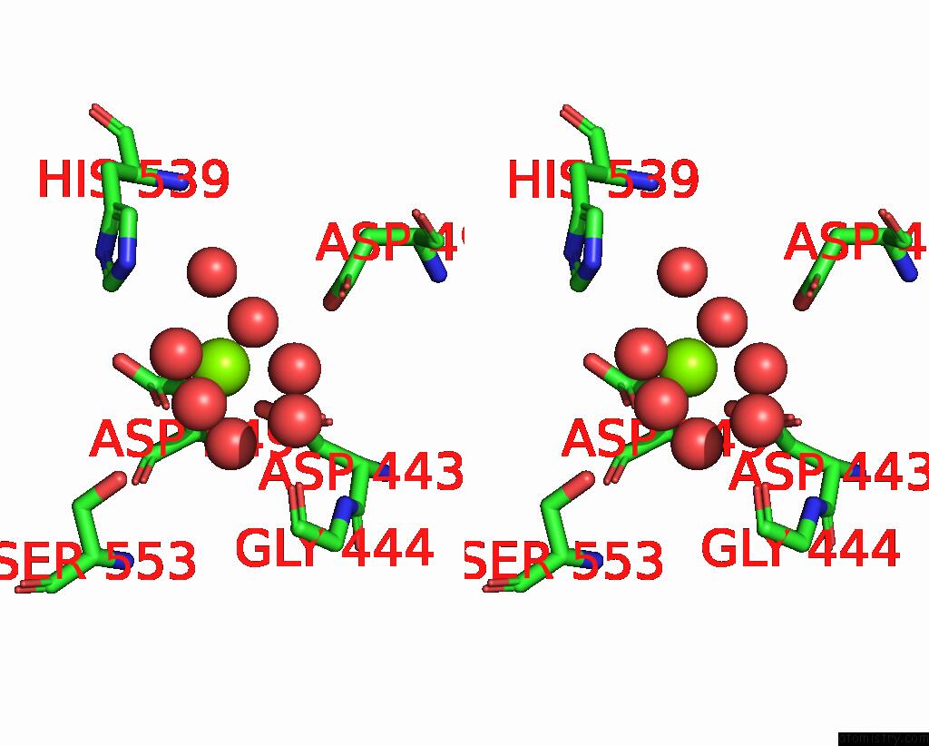
Stereo pair view
Mono view
Stereo pair view
A full contact list of Magnesium with other atoms in the Mg binding
site number 1 of Hiv-1 Reverse Transcriptase/Rilpivirine with Bound Fragment 4- Iodopyrazole at Multiple Sites within 5.0Å range:
|
Reference:
A.Chopra,
J.D.Bauman,
F.X.Ruiz,
E.Arnold.
Halo Library, A Tool For Rapid Identification of Ligand Binding Sites on Proteins Using Crystallographic Fragment Screening. J.Med.Chem. V. 66 6013 2023.
ISSN: ISSN 0022-2623
PubMed: 37115705
DOI: 10.1021/ACS.JMEDCHEM.2C01681
Page generated: Fri Oct 4 00:58:26 2024
ISSN: ISSN 0022-2623
PubMed: 37115705
DOI: 10.1021/ACS.JMEDCHEM.2C01681
Last articles
Fe in 2YXOFe in 2YRS
Fe in 2YXC
Fe in 2YNM
Fe in 2YVJ
Fe in 2YP1
Fe in 2YU2
Fe in 2YU1
Fe in 2YQB
Fe in 2YOO