Magnesium »
PDB 8ezg-8f8q »
8f2w »
Magnesium in PDB 8f2w: Structure of A B-Form Dodecamer: 5'-Cgcgaattcgcg-3
Protein crystallography data
The structure of Structure of A B-Form Dodecamer: 5'-Cgcgaattcgcg-3, PDB code: 8f2w
was solved by
E.N.Ogbonna,
W.D.Wilson,
with X-Ray Crystallography technique. A brief refinement statistics is given in the table below:
| Resolution Low / High (Å) | 34.29 / 1.30 |
| Space group | P 21 21 21 |
| Cell size a, b, c (Å), α, β, γ (°) | 25.58, 40.104, 66.099, 90, 90, 90 |
| R / Rfree (%) | 18.7 / 21.5 |
Magnesium Binding Sites:
The binding sites of Magnesium atom in the Structure of A B-Form Dodecamer: 5'-Cgcgaattcgcg-3
(pdb code 8f2w). This binding sites where shown within
5.0 Angstroms radius around Magnesium atom.
In total only one binding site of Magnesium was determined in the Structure of A B-Form Dodecamer: 5'-Cgcgaattcgcg-3, PDB code: 8f2w:
In total only one binding site of Magnesium was determined in the Structure of A B-Form Dodecamer: 5'-Cgcgaattcgcg-3, PDB code: 8f2w:
Magnesium binding site 1 out of 1 in 8f2w
Go back to
Magnesium binding site 1 out
of 1 in the Structure of A B-Form Dodecamer: 5'-Cgcgaattcgcg-3

Mono view
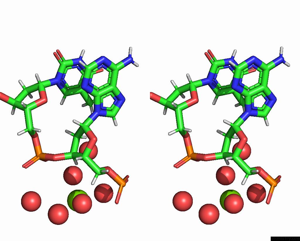
Stereo pair view
Mono view
Stereo pair view
A full contact list of Magnesium with other atoms in the Mg binding
site number 1 of Structure of A B-Form Dodecamer: 5'-Cgcgaattcgcg-3 within 5.0Å range:
|
Reference:
E.N.Ogbonna,
A.Paul,
A.Faharat,
J.R.Terrell,
V.Ogbonna,
D.W.Boykin,
W.D.Wilson.
X-Ray Structure Characterization of the Selective Recognition of at Base Pair Sequences Acs Bio Med Chem Au 2023.
ISSN: ESSN 2694-2437
Page generated: Fri Aug 15 04:02:22 2025
ISSN: ESSN 2694-2437
Last articles
Mg in 8HCCMg in 8HCH
Mg in 8HCG
Mg in 8HBH
Mg in 8HB1
Mg in 8HBF
Mg in 8H6U
Mg in 8H9V
Mg in 8HAC
Mg in 8H99