Magnesium »
PDB 8rav-8rjw »
8re2 »
Magnesium in PDB 8re2: Crystal Structure Determination of Dye-Decolorizing Peroxidase (Dyp) From Deinoccoccus Radiodurans
Protein crystallography data
The structure of Crystal Structure Determination of Dye-Decolorizing Peroxidase (Dyp) From Deinoccoccus Radiodurans, PDB code: 8re2
was solved by
B.A.Salgueiro,
K.Frade,
C.Frazao,
E.Moe,
with X-Ray Crystallography technique. A brief refinement statistics is given in the table below:
| Resolution Low / High (Å) | 39.31 / 2.20 |
| Space group | P 32 |
| Cell size a, b, c (Å), α, β, γ (°) | 64.13, 64.13, 111.322, 90, 90, 120 |
| R / Rfree (%) | 18.3 / 23.7 |
Other elements in 8re2:
The structure of Crystal Structure Determination of Dye-Decolorizing Peroxidase (Dyp) From Deinoccoccus Radiodurans also contains other interesting chemical elements:
| Iron | (Fe) | 1 atom |
Magnesium Binding Sites:
The binding sites of Magnesium atom in the Crystal Structure Determination of Dye-Decolorizing Peroxidase (Dyp) From Deinoccoccus Radiodurans
(pdb code 8re2). This binding sites where shown within
5.0 Angstroms radius around Magnesium atom.
In total 3 binding sites of Magnesium where determined in the Crystal Structure Determination of Dye-Decolorizing Peroxidase (Dyp) From Deinoccoccus Radiodurans, PDB code: 8re2:
Jump to Magnesium binding site number: 1; 2; 3;
In total 3 binding sites of Magnesium where determined in the Crystal Structure Determination of Dye-Decolorizing Peroxidase (Dyp) From Deinoccoccus Radiodurans, PDB code: 8re2:
Jump to Magnesium binding site number: 1; 2; 3;
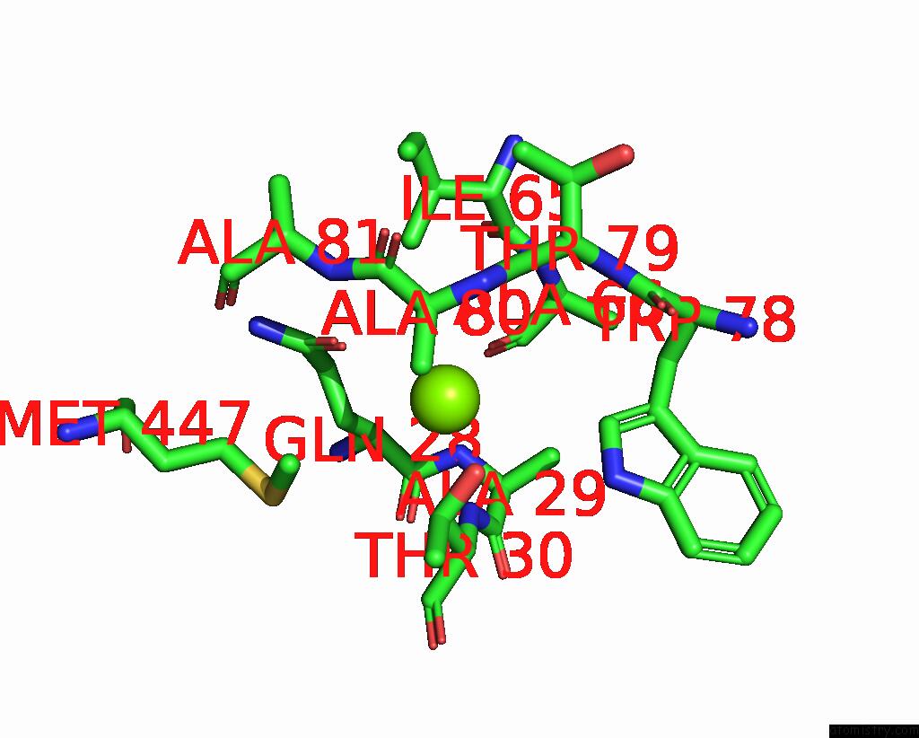
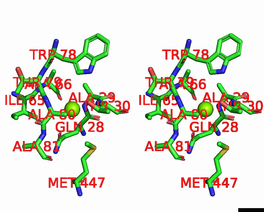
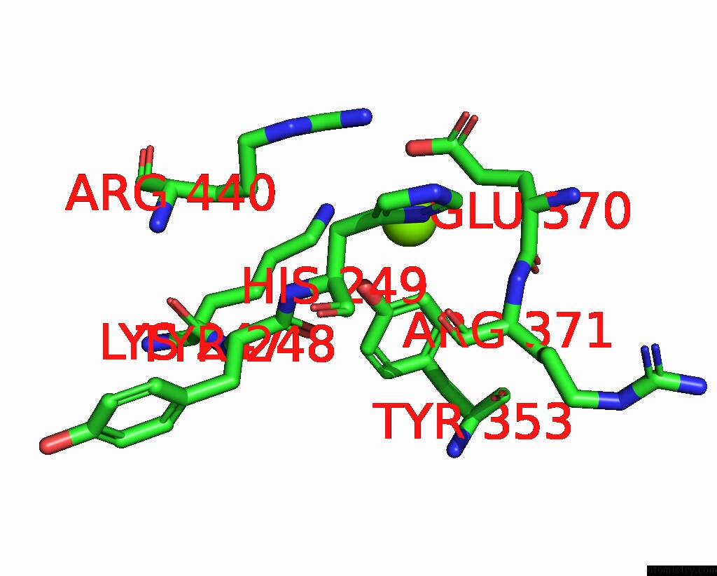
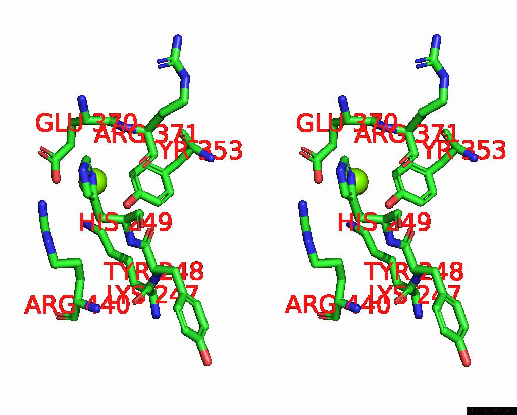
Magnesium binding site 1 out of 3 in 8re2
Go back to
Magnesium binding site 1 out
of 3 in the Crystal Structure Determination of Dye-Decolorizing Peroxidase (Dyp) From Deinoccoccus Radiodurans
Mono view
Stereo pair view
Mono view
Stereo pair view
A full contact list of Magnesium with other atoms in the Mg binding
site number 1 of Crystal Structure Determination of Dye-Decolorizing Peroxidase (Dyp) From Deinoccoccus Radiodurans within 5.0Å range:
|
Magnesium binding site 2 out of 3 in 8re2
Go back to
Magnesium binding site 2 out
of 3 in the Crystal Structure Determination of Dye-Decolorizing Peroxidase (Dyp) From Deinoccoccus Radiodurans
Mono view
Stereo pair view
Mono view
Stereo pair view
A full contact list of Magnesium with other atoms in the Mg binding
site number 2 of Crystal Structure Determination of Dye-Decolorizing Peroxidase (Dyp) From Deinoccoccus Radiodurans within 5.0Å range:
|
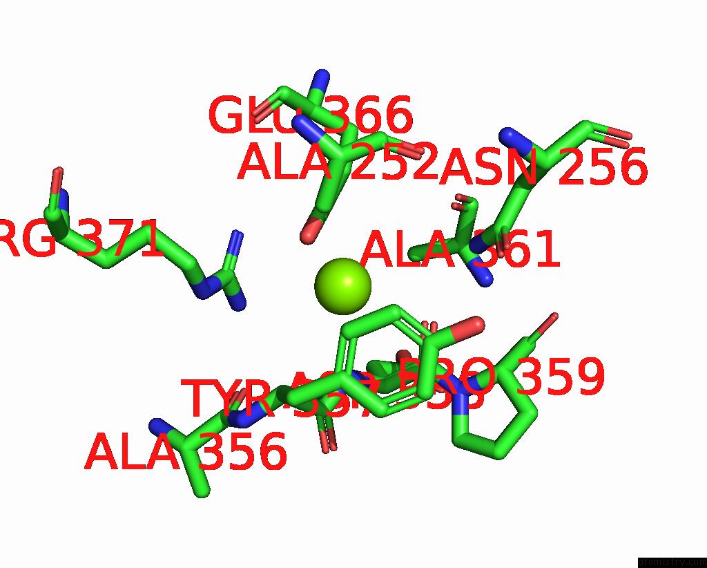
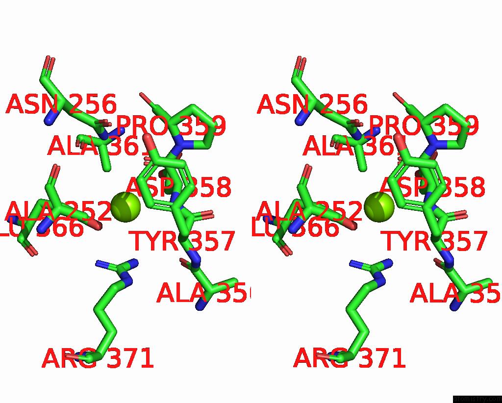
Magnesium binding site 3 out of 3 in 8re2
Go back to
Magnesium binding site 3 out
of 3 in the Crystal Structure Determination of Dye-Decolorizing Peroxidase (Dyp) From Deinoccoccus Radiodurans
Mono view
Stereo pair view
Mono view
Stereo pair view
A full contact list of Magnesium with other atoms in the Mg binding
site number 3 of Crystal Structure Determination of Dye-Decolorizing Peroxidase (Dyp) From Deinoccoccus Radiodurans within 5.0Å range:
|
Reference:
K.Frade,
C.M.Silveira,
B.A.Salgueiro,
S.Mendes,
L.O.Martins,
C.Frazao,
S.Todorovic,
E.Moe.
Biochemical, Biophysical, and Structural Analysis of An Unusual Dyp From the Extremophile Deinococcus Radiodurans. Molecules V. 29 2024.
ISSN: ESSN 1420-3049
PubMed: 38257271
DOI: 10.3390/MOLECULES29020358
Page generated: Fri Aug 15 13:57:17 2025
ISSN: ESSN 1420-3049
PubMed: 38257271
DOI: 10.3390/MOLECULES29020358
Last articles
Mg in 8UV4Mg in 8UVA
Mg in 8UV9
Mg in 8UV8
Mg in 8UV3
Mg in 8UTY
Mg in 8UUC
Mg in 8UPP
Mg in 8UTW
Mg in 8UTT