Magnesium »
PDB 8shf-8srd »
8slf »
Magnesium in PDB 8slf: Crystal Structure of Glycine Trna Ligase From Mycobacterium Thermoresistibile (Amp Bound)
Protein crystallography data
The structure of Crystal Structure of Glycine Trna Ligase From Mycobacterium Thermoresistibile (Amp Bound), PDB code: 8slf
was solved by
Seattle Structural Genomics Center For Infectious Disease (Ssgcid),
with X-Ray Crystallography technique. A brief refinement statistics is given in the table below:
| Resolution Low / High (Å) | 42.60 / 2.90 |
| Space group | C 1 2 1 |
| Cell size a, b, c (Å), α, β, γ (°) | 173.757, 86.297, 98.986, 90, 104.2, 90 |
| R / Rfree (%) | 18.3 / 22.4 |
Other elements in 8slf:
The structure of Crystal Structure of Glycine Trna Ligase From Mycobacterium Thermoresistibile (Amp Bound) also contains other interesting chemical elements:
| Zinc | (Zn) | 1 atom |
Magnesium Binding Sites:
The binding sites of Magnesium atom in the Crystal Structure of Glycine Trna Ligase From Mycobacterium Thermoresistibile (Amp Bound)
(pdb code 8slf). This binding sites where shown within
5.0 Angstroms radius around Magnesium atom.
In total 2 binding sites of Magnesium where determined in the Crystal Structure of Glycine Trna Ligase From Mycobacterium Thermoresistibile (Amp Bound), PDB code: 8slf:
Jump to Magnesium binding site number: 1; 2;
In total 2 binding sites of Magnesium where determined in the Crystal Structure of Glycine Trna Ligase From Mycobacterium Thermoresistibile (Amp Bound), PDB code: 8slf:
Jump to Magnesium binding site number: 1; 2;
Magnesium binding site 1 out of 2 in 8slf
Go back to
Magnesium binding site 1 out
of 2 in the Crystal Structure of Glycine Trna Ligase From Mycobacterium Thermoresistibile (Amp Bound)
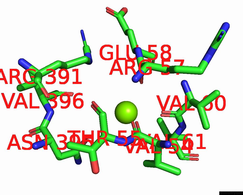
Mono view
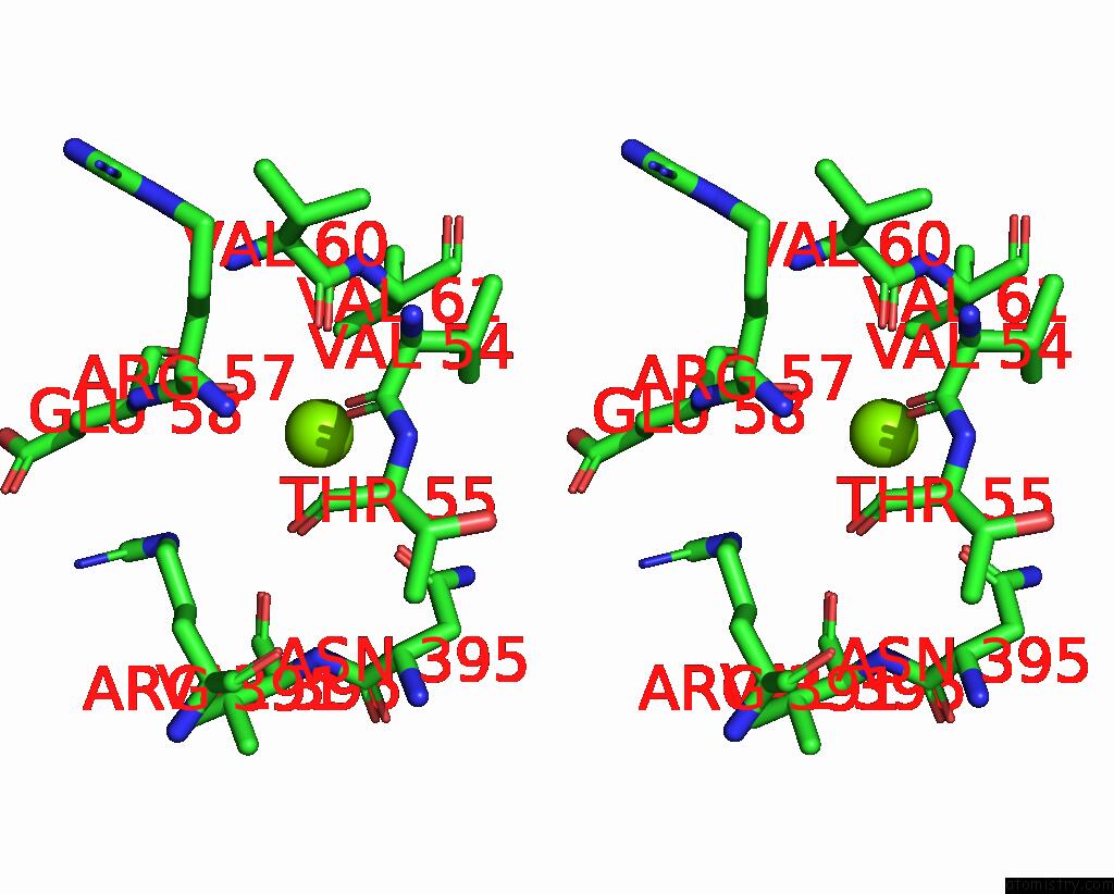
Stereo pair view
Mono view
Stereo pair view
A full contact list of Magnesium with other atoms in the Mg binding
site number 1 of Crystal Structure of Glycine Trna Ligase From Mycobacterium Thermoresistibile (Amp Bound) within 5.0Å range:
|
Magnesium binding site 2 out of 2 in 8slf
Go back to
Magnesium binding site 2 out
of 2 in the Crystal Structure of Glycine Trna Ligase From Mycobacterium Thermoresistibile (Amp Bound)
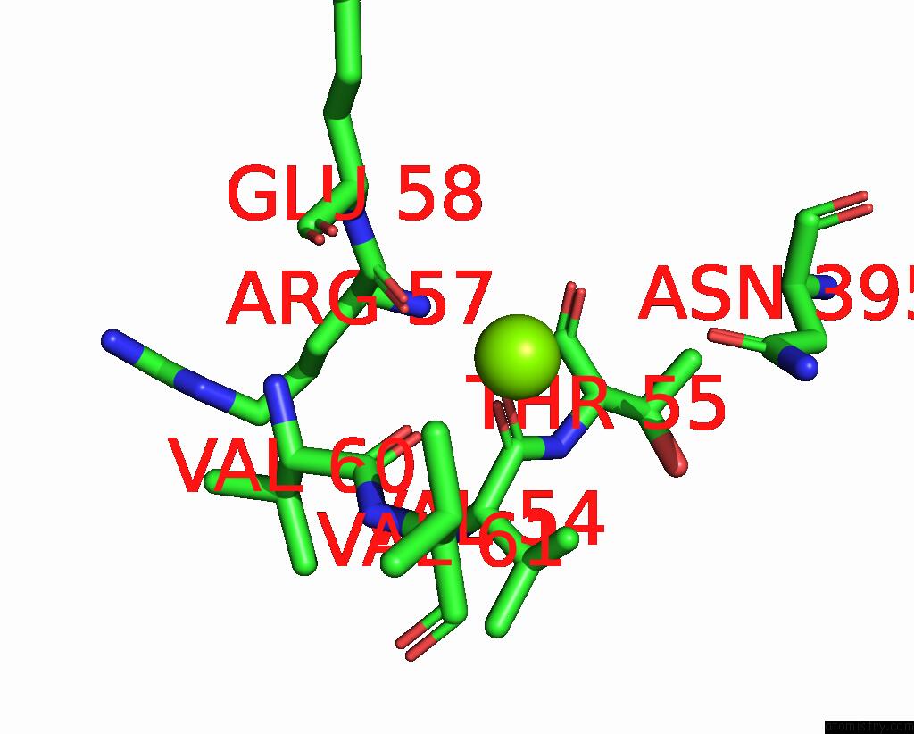
Mono view

Stereo pair view
Mono view
Stereo pair view
A full contact list of Magnesium with other atoms in the Mg binding
site number 2 of Crystal Structure of Glycine Trna Ligase From Mycobacterium Thermoresistibile (Amp Bound) within 5.0Å range:
|
Reference:
S.Lovell,
L.Liu,
K.P.Battaile,
S.Seibold.
Crystal Structure of Glycine Trna Ligase From Mycobacterium Thermoresistibile (Amp Bound) To Be Published.
Page generated: Fri Aug 15 15:47:40 2025
Last articles
Mg in 8WCKMg in 8WCE
Mg in 8WB0
Mg in 8WB1
Mg in 8WAY
Mg in 8WAZ
Mg in 8WAX
Mg in 8WAV
Mg in 8WAW
Mg in 8WAU