Magnesium »
PDB 8shf-8srd »
8smf »
Magnesium in PDB 8smf: Structure of SPO1 Phage TAD2 in Complex with 1''-3' Gcadpr
Protein crystallography data
The structure of Structure of SPO1 Phage TAD2 in Complex with 1''-3' Gcadpr, PDB code: 8smf
was solved by
A.Lu,
E.Yirmiya,
A.Leavitt,
C.Avraham,
I.Osterman,
J.Garb,
S.P.Antine,
S.E.Mooney,
S.J.Hobbs,
G.Amitai,
R.Sorek,
P.J.Kranzusch,
with X-Ray Crystallography technique. A brief refinement statistics is given in the table below:
| Resolution Low / High (Å) | 46.06 / 1.75 |
| Space group | C 1 2 1 |
| Cell size a, b, c (Å), α, β, γ (°) | 94.389, 82.472, 90.757, 90, 107.88, 90 |
| R / Rfree (%) | 20.8 / 24.1 |
Magnesium Binding Sites:
The binding sites of Magnesium atom in the Structure of SPO1 Phage TAD2 in Complex with 1''-3' Gcadpr
(pdb code 8smf). This binding sites where shown within
5.0 Angstroms radius around Magnesium atom.
In total 2 binding sites of Magnesium where determined in the Structure of SPO1 Phage TAD2 in Complex with 1''-3' Gcadpr, PDB code: 8smf:
Jump to Magnesium binding site number: 1; 2;
In total 2 binding sites of Magnesium where determined in the Structure of SPO1 Phage TAD2 in Complex with 1''-3' Gcadpr, PDB code: 8smf:
Jump to Magnesium binding site number: 1; 2;
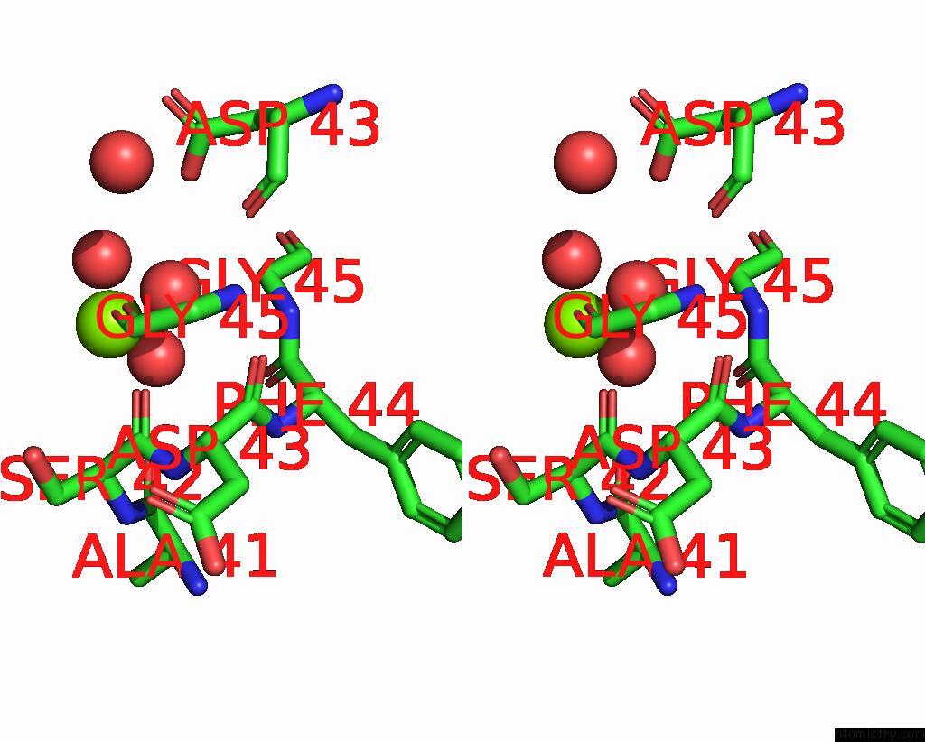
Magnesium binding site 1 out of 2 in 8smf
Go back to
Magnesium binding site 1 out
of 2 in the Structure of SPO1 Phage TAD2 in Complex with 1''-3' Gcadpr

Mono view
Stereo pair view
Mono view
Stereo pair view
A full contact list of Magnesium with other atoms in the Mg binding
site number 1 of Structure of SPO1 Phage TAD2 in Complex with 1''-3' Gcadpr within 5.0Å range:
|
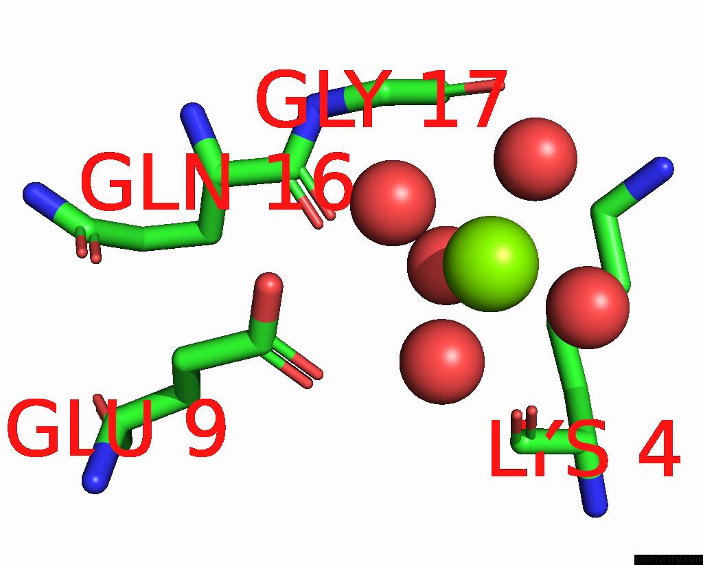
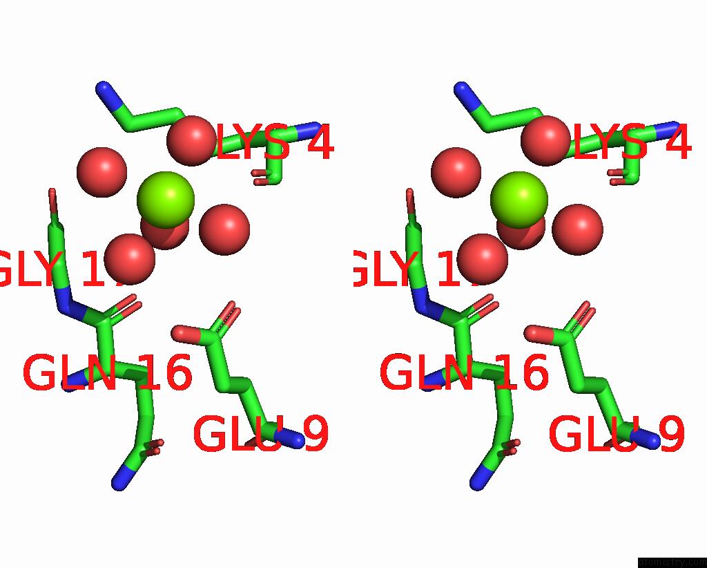
Magnesium binding site 2 out of 2 in 8smf
Go back to
Magnesium binding site 2 out
of 2 in the Structure of SPO1 Phage TAD2 in Complex with 1''-3' Gcadpr
Mono view
Stereo pair view
Mono view
Stereo pair view
A full contact list of Magnesium with other atoms in the Mg binding
site number 2 of Structure of SPO1 Phage TAD2 in Complex with 1''-3' Gcadpr within 5.0Å range:
|
Reference:
E.Yirmiya,
A.Leavitt,
A.Lu,
C.Avraham,
I.Osterman,
J.Garb,
S.P.Antine,
S.E.Mooney,
S.J.Hobbs,
P.J.Kranzusch,
G.Amitai,
R.Sorek.
Phages Overcome Bacterial Immunity Via Diverse Anti-Defense Proteins To Be Published.
Page generated: Fri Aug 15 15:48:11 2025
Last articles
Mg in 8WCKMg in 8WCE
Mg in 8WB0
Mg in 8WB1
Mg in 8WAY
Mg in 8WAZ
Mg in 8WAX
Mg in 8WAV
Mg in 8WAW
Mg in 8WAU