Magnesium »
PDB 1s9d-1so5 »
1sh3 »
Magnesium in PDB 1sh3: Crystal Structure of Norwalk Virus Polymerase (MGSO4 Crystal Form)
Enzymatic activity of Crystal Structure of Norwalk Virus Polymerase (MGSO4 Crystal Form)
All present enzymatic activity of Crystal Structure of Norwalk Virus Polymerase (MGSO4 Crystal Form):
2.7.7.48;
2.7.7.48;
Protein crystallography data
The structure of Crystal Structure of Norwalk Virus Polymerase (MGSO4 Crystal Form), PDB code: 1sh3
was solved by
K.K.Ng,
N.Pendas-Franco,
J.Rojo,
J.A.Boga,
A.Machin,
J.M.Alonso,
F.Parra,
with X-Ray Crystallography technique. A brief refinement statistics is given in the table below:
| Resolution Low / High (Å) | 20.00 / 2.95 |
| Space group | P 21 21 21 |
| Cell size a, b, c (Å), α, β, γ (°) | 105.050, 109.140, 112.044, 90.00, 90.00, 90.00 |
| R / Rfree (%) | 21.5 / 28 |
Magnesium Binding Sites:
The binding sites of Magnesium atom in the Crystal Structure of Norwalk Virus Polymerase (MGSO4 Crystal Form)
(pdb code 1sh3). This binding sites where shown within
5.0 Angstroms radius around Magnesium atom.
In total 2 binding sites of Magnesium where determined in the Crystal Structure of Norwalk Virus Polymerase (MGSO4 Crystal Form), PDB code: 1sh3:
Jump to Magnesium binding site number: 1; 2;
In total 2 binding sites of Magnesium where determined in the Crystal Structure of Norwalk Virus Polymerase (MGSO4 Crystal Form), PDB code: 1sh3:
Jump to Magnesium binding site number: 1; 2;
Magnesium binding site 1 out of 2 in 1sh3
Go back to
Magnesium binding site 1 out
of 2 in the Crystal Structure of Norwalk Virus Polymerase (MGSO4 Crystal Form)
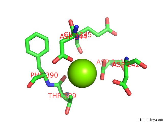
Mono view
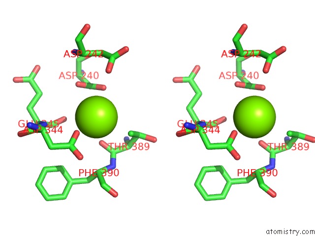
Stereo pair view
Mono view
Stereo pair view
A full contact list of Magnesium with other atoms in the Mg binding
site number 1 of Crystal Structure of Norwalk Virus Polymerase (MGSO4 Crystal Form) within 5.0Å range:
|
Magnesium binding site 2 out of 2 in 1sh3
Go back to
Magnesium binding site 2 out
of 2 in the Crystal Structure of Norwalk Virus Polymerase (MGSO4 Crystal Form)
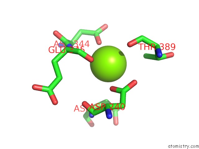
Mono view
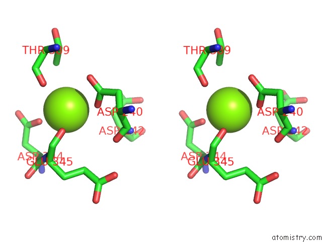
Stereo pair view
Mono view
Stereo pair view
A full contact list of Magnesium with other atoms in the Mg binding
site number 2 of Crystal Structure of Norwalk Virus Polymerase (MGSO4 Crystal Form) within 5.0Å range:
|
Reference:
K.K.Ng,
N.Pendas-Franco,
J.Rojo,
J.A.Boga,
A.Machin,
J.M.Alonso,
F.Parra.
Crystal Structure of Norwalk Virus Polymerase Reveals the Carboxyl Terminus in the Active Site Cleft. J.Biol.Chem. V. 279 16638 2004.
ISSN: ISSN 0021-9258
PubMed: 14764591
DOI: 10.1074/JBC.M400584200
Page generated: Sun Aug 10 04:22:15 2025
ISSN: ISSN 0021-9258
PubMed: 14764591
DOI: 10.1074/JBC.M400584200
Last articles
Mg in 4AV3Mg in 4AUX
Mg in 4ATB
Mg in 4AUI
Mg in 4AT9
Mg in 4AT8
Mg in 4AS5
Mg in 4ASU
Mg in 4AS8
Mg in 4AS4