Magnesium »
PDB 1so6-1t5s »
1t1s »
Magnesium in PDB 1t1s: Crystal Structure of the Reductoisomerase Complexed with A Bisphosphonate
Enzymatic activity of Crystal Structure of the Reductoisomerase Complexed with A Bisphosphonate
All present enzymatic activity of Crystal Structure of the Reductoisomerase Complexed with A Bisphosphonate:
1.1.1.267;
1.1.1.267;
Protein crystallography data
The structure of Crystal Structure of the Reductoisomerase Complexed with A Bisphosphonate, PDB code: 1t1s
was solved by
S.Yajima,
K.Hara,
J.M.Sanders,
F.Yin,
K.Ohsawa,
J.Wiesner,
H.Jomaa,
E.Oldfield,
with X-Ray Crystallography technique. A brief refinement statistics is given in the table below:
| Resolution Low / High (Å) | 50.00 / 2.40 |
| Space group | P 21 21 2 |
| Cell size a, b, c (Å), α, β, γ (°) | 182.917, 59.246, 86.988, 90.00, 90.00, 90.00 |
| R / Rfree (%) | 21.3 / 26 |
Other elements in 1t1s:
The structure of Crystal Structure of the Reductoisomerase Complexed with A Bisphosphonate also contains other interesting chemical elements:
| Chlorine | (Cl) | 2 atoms |
Magnesium Binding Sites:
The binding sites of Magnesium atom in the Crystal Structure of the Reductoisomerase Complexed with A Bisphosphonate
(pdb code 1t1s). This binding sites where shown within
5.0 Angstroms radius around Magnesium atom.
In total only one binding site of Magnesium was determined in the Crystal Structure of the Reductoisomerase Complexed with A Bisphosphonate, PDB code: 1t1s:
In total only one binding site of Magnesium was determined in the Crystal Structure of the Reductoisomerase Complexed with A Bisphosphonate, PDB code: 1t1s:
Magnesium binding site 1 out of 1 in 1t1s
Go back to
Magnesium binding site 1 out
of 1 in the Crystal Structure of the Reductoisomerase Complexed with A Bisphosphonate
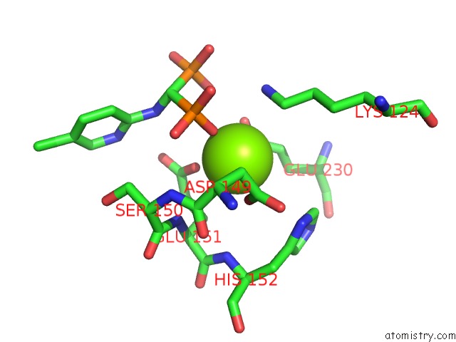
Mono view
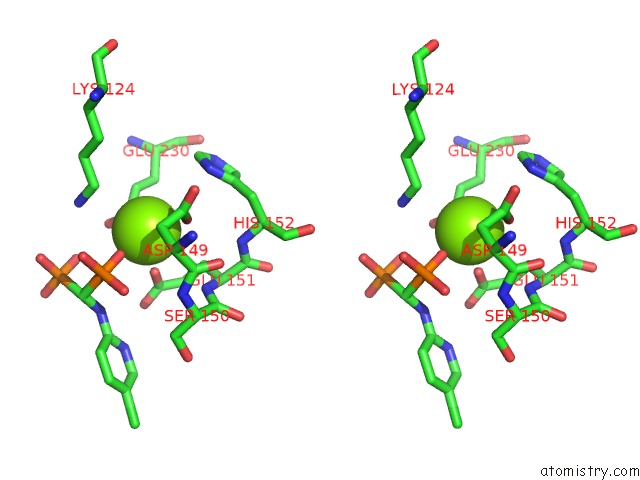
Stereo pair view
Mono view
Stereo pair view
A full contact list of Magnesium with other atoms in the Mg binding
site number 1 of Crystal Structure of the Reductoisomerase Complexed with A Bisphosphonate within 5.0Å range:
|
Reference:
S.Yajima,
K.Hara,
J.M.Sanders,
F.Yin,
K.Ohsawa,
J.Wiesner,
H.Jomaa,
E.Oldfield.
Crystallographic Structures of Two Bisphosphonate:1-Deoxyxylulose-5-Phosphate Reductoisomerase Complexes J.Am.Chem.Soc. V. 126 10824 2004.
ISSN: ISSN 0002-7863
PubMed: 15339150
DOI: 10.1021/JA040126M
Page generated: Tue Aug 13 14:25:00 2024
ISSN: ISSN 0002-7863
PubMed: 15339150
DOI: 10.1021/JA040126M
Last articles
Mg in 1RYPMg in 1S2O
Mg in 1S0V
Mg in 1S1C
Mg in 1S16
Mg in 1S0P
Mg in 1S0O
Mg in 1S0M
Mg in 1RZZ
Mg in 1S00