Magnesium »
PDB 2amc-2b2k »
2b0c »
Magnesium in PDB 2b0c: The Crystal Structure of the Putative Phosphatase From Escherichia Coli
Protein crystallography data
The structure of The Crystal Structure of the Putative Phosphatase From Escherichia Coli, PDB code: 2b0c
was solved by
R.Zhang,
T.Skarina,
A.Savchenko,
A.Edwards,
A.Joachimiak,
Midwest Centerfor Structural Genomics (Mcsg),
with X-Ray Crystallography technique. A brief refinement statistics is given in the table below:
| Resolution Low / High (Å) | 50.00 / 2.00 |
| Space group | P 41 21 2 |
| Cell size a, b, c (Å), α, β, γ (°) | 62.256, 62.256, 127.038, 90.00, 90.00, 90.00 |
| R / Rfree (%) | 20.1 / 25.9 |
Magnesium Binding Sites:
The binding sites of Magnesium atom in the The Crystal Structure of the Putative Phosphatase From Escherichia Coli
(pdb code 2b0c). This binding sites where shown within
5.0 Angstroms radius around Magnesium atom.
In total only one binding site of Magnesium was determined in the The Crystal Structure of the Putative Phosphatase From Escherichia Coli, PDB code: 2b0c:
In total only one binding site of Magnesium was determined in the The Crystal Structure of the Putative Phosphatase From Escherichia Coli, PDB code: 2b0c:
Magnesium binding site 1 out of 1 in 2b0c
Go back to
Magnesium binding site 1 out
of 1 in the The Crystal Structure of the Putative Phosphatase From Escherichia Coli
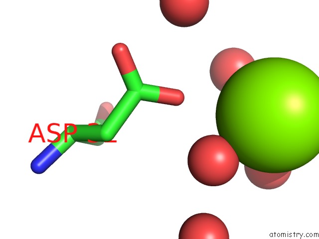
Mono view
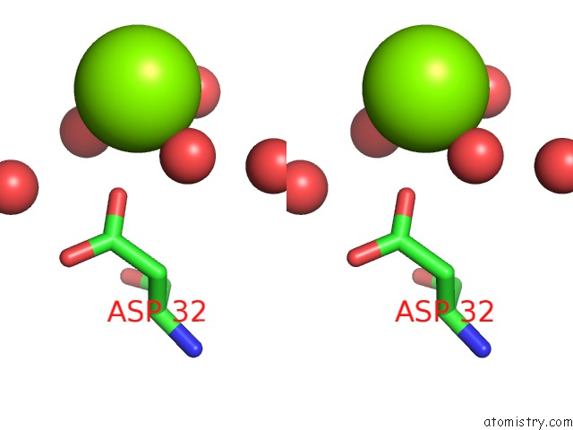
Stereo pair view
Mono view
Stereo pair view
A full contact list of Magnesium with other atoms in the Mg binding
site number 1 of The Crystal Structure of the Putative Phosphatase From Escherichia Coli within 5.0Å range:
|
Reference:
R.Zhang,
T.Skarina,
A.Savchenko,
A.Edwards,
A.Joachimiak.
The 2.0A Crystal Structure of the Putative Phosphatase From Escherichia Coli To Be Published.
Page generated: Sun Aug 10 09:51:40 2025
Last articles
Mg in 5WMBMg in 5WM8
Mg in 5WNO
Mg in 5WNI
Mg in 5WMT
Mg in 5WM1
Mg in 5WM6
Mg in 5WM4
Mg in 5WKC
Mg in 5WM3