Magnesium »
PDB 2gqr-2haw »
2gt4 »
Magnesium in PDB 2gt4: Crystal Structure of the Y103F Mutant of the Gdp-Mannose Mannosyl Hydrolase in Complex with Gdp-Mannose and Mg+2
Protein crystallography data
The structure of Crystal Structure of the Y103F Mutant of the Gdp-Mannose Mannosyl Hydrolase in Complex with Gdp-Mannose and Mg+2, PDB code: 2gt4
was solved by
S.B.Gabelli,
M.A.Bianchet,
H.F.Azurmendi,
A.S.Mildvan,
L.A.Amzel,
with X-Ray Crystallography technique. A brief refinement statistics is given in the table below:
| Resolution Low / High (Å) | 77.61 / 2.30 |
| Space group | C 1 2 1 |
| Cell size a, b, c (Å), α, β, γ (°) | 137.785, 93.893, 66.103, 90.00, 91.23, 90.00 |
| R / Rfree (%) | 18.6 / 22.6 |
Magnesium Binding Sites:
The binding sites of Magnesium atom in the Crystal Structure of the Y103F Mutant of the Gdp-Mannose Mannosyl Hydrolase in Complex with Gdp-Mannose and Mg+2
(pdb code 2gt4). This binding sites where shown within
5.0 Angstroms radius around Magnesium atom.
In total 3 binding sites of Magnesium where determined in the Crystal Structure of the Y103F Mutant of the Gdp-Mannose Mannosyl Hydrolase in Complex with Gdp-Mannose and Mg+2, PDB code: 2gt4:
Jump to Magnesium binding site number: 1; 2; 3;
In total 3 binding sites of Magnesium where determined in the Crystal Structure of the Y103F Mutant of the Gdp-Mannose Mannosyl Hydrolase in Complex with Gdp-Mannose and Mg+2, PDB code: 2gt4:
Jump to Magnesium binding site number: 1; 2; 3;
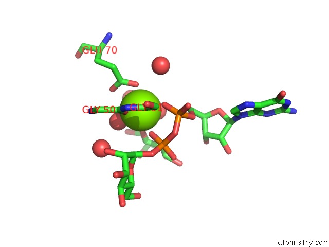
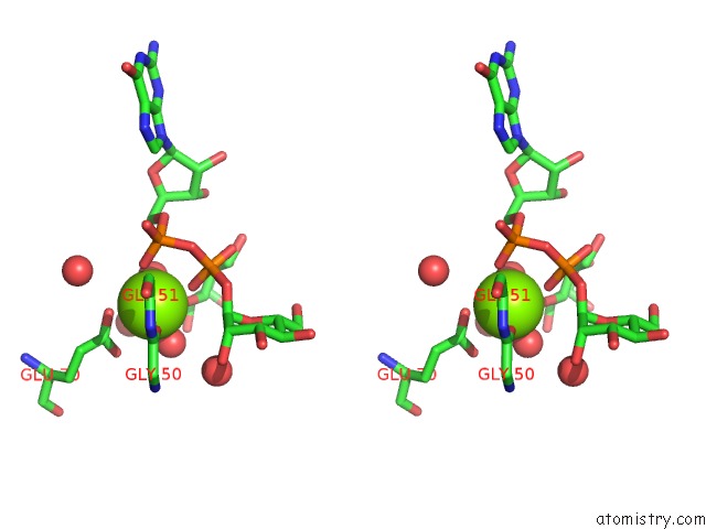
Magnesium binding site 1 out of 3 in 2gt4
Go back to
Magnesium binding site 1 out
of 3 in the Crystal Structure of the Y103F Mutant of the Gdp-Mannose Mannosyl Hydrolase in Complex with Gdp-Mannose and Mg+2
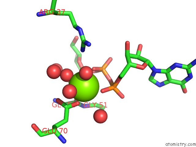
Mono view
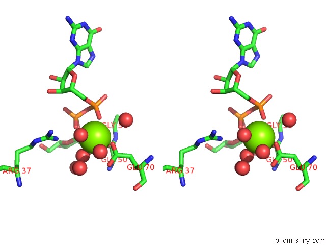
Stereo pair view
Mono view
Stereo pair view
A full contact list of Magnesium with other atoms in the Mg binding
site number 1 of Crystal Structure of the Y103F Mutant of the Gdp-Mannose Mannosyl Hydrolase in Complex with Gdp-Mannose and Mg+2 within 5.0Å range:
|
Magnesium binding site 2 out of 3 in 2gt4
Go back to
Magnesium binding site 2 out
of 3 in the Crystal Structure of the Y103F Mutant of the Gdp-Mannose Mannosyl Hydrolase in Complex with Gdp-Mannose and Mg+2
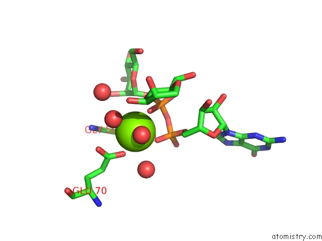
Mono view
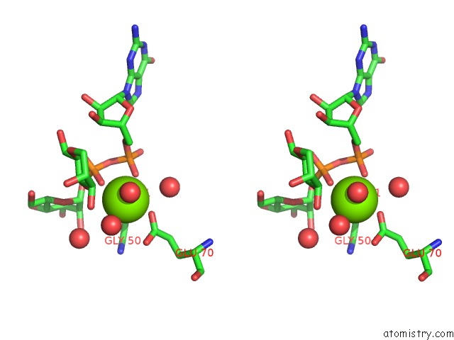
Stereo pair view
Mono view
Stereo pair view
A full contact list of Magnesium with other atoms in the Mg binding
site number 2 of Crystal Structure of the Y103F Mutant of the Gdp-Mannose Mannosyl Hydrolase in Complex with Gdp-Mannose and Mg+2 within 5.0Å range:
|
Magnesium binding site 3 out of 3 in 2gt4
Go back to
Magnesium binding site 3 out
of 3 in the Crystal Structure of the Y103F Mutant of the Gdp-Mannose Mannosyl Hydrolase in Complex with Gdp-Mannose and Mg+2
Mono view
Stereo pair view
Mono view
Stereo pair view
A full contact list of Magnesium with other atoms in the Mg binding
site number 3 of Crystal Structure of the Y103F Mutant of the Gdp-Mannose Mannosyl Hydrolase in Complex with Gdp-Mannose and Mg+2 within 5.0Å range:
|
Reference:
S.B.Gabelli,
H.F.Azurmendi,
M.A.Bianchet,
L.M.Amzel,
A.S.Mildvan.
X-Ray, uc(Nmr), and Mutational Studies of the Catalytic Cycle of the Gdp-Mannose Mannosyl Hydrolase Reaction. Biochemistry V. 45 11290 2006.
ISSN: ISSN 0006-2960
PubMed: 16981689
DOI: 10.1021/BI061239G
Page generated: Sun Aug 10 11:11:34 2025
ISSN: ISSN 0006-2960
PubMed: 16981689
DOI: 10.1021/BI061239G
Last articles
Mg in 5L3SMg in 5L44
Mg in 5L3R
Mg in 5L43
Mg in 5L3Q
Mg in 5L22
Mg in 5L1L
Mg in 5L1K
Mg in 5L1J
Mg in 5L0Q