Magnesium »
PDB 2wkp-2x0q »
2x03 »
Magnesium in PDB 2x03: The X-Ray Structure of the Streptomyces Coelicolor A3 Chondroitin Ac Lyase Y253A Mutant
Enzymatic activity of The X-Ray Structure of the Streptomyces Coelicolor A3 Chondroitin Ac Lyase Y253A Mutant
All present enzymatic activity of The X-Ray Structure of the Streptomyces Coelicolor A3 Chondroitin Ac Lyase Y253A Mutant:
4.2.2.1;
4.2.2.1;
Protein crystallography data
The structure of The X-Ray Structure of the Streptomyces Coelicolor A3 Chondroitin Ac Lyase Y253A Mutant, PDB code: 2x03
was solved by
Z.H.Elmabrouk,
E.J.Taylor,
F.Vincent,
N.L.Smith,
J.P.Turkenburg,
G.J.Davies,
G.W.Black,
with X-Ray Crystallography technique. A brief refinement statistics is given in the table below:
| Resolution Low / High (Å) | 158.55 / 2.30 |
| Space group | H 3 |
| Cell size a, b, c (Å), α, β, γ (°) | 317.095, 317.095, 82.976, 90.00, 90.00, 120.00 |
| R / Rfree (%) | 19.4 / 22.8 |
Magnesium Binding Sites:
The binding sites of Magnesium atom in the The X-Ray Structure of the Streptomyces Coelicolor A3 Chondroitin Ac Lyase Y253A Mutant
(pdb code 2x03). This binding sites where shown within
5.0 Angstroms radius around Magnesium atom.
In total only one binding site of Magnesium was determined in the The X-Ray Structure of the Streptomyces Coelicolor A3 Chondroitin Ac Lyase Y253A Mutant, PDB code: 2x03:
In total only one binding site of Magnesium was determined in the The X-Ray Structure of the Streptomyces Coelicolor A3 Chondroitin Ac Lyase Y253A Mutant, PDB code: 2x03:
Magnesium binding site 1 out of 1 in 2x03
Go back to
Magnesium binding site 1 out
of 1 in the The X-Ray Structure of the Streptomyces Coelicolor A3 Chondroitin Ac Lyase Y253A Mutant
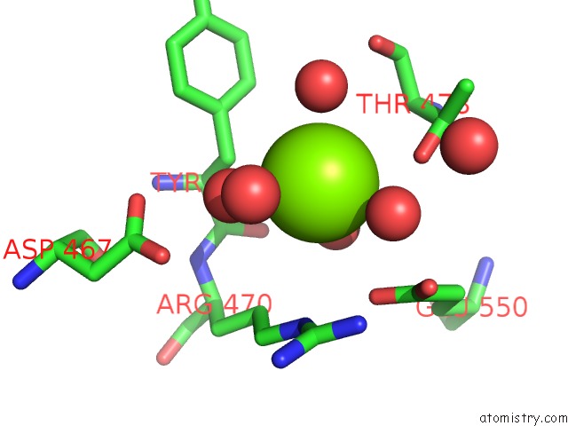
Mono view
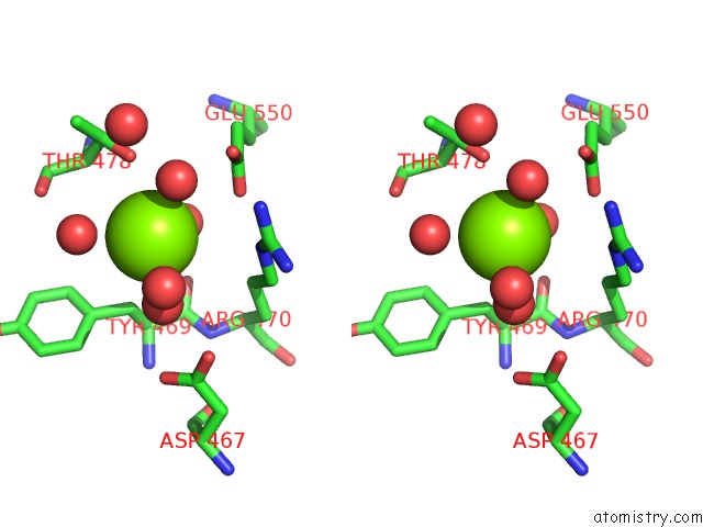
Stereo pair view
Mono view
Stereo pair view
A full contact list of Magnesium with other atoms in the Mg binding
site number 1 of The X-Ray Structure of the Streptomyces Coelicolor A3 Chondroitin Ac Lyase Y253A Mutant within 5.0Å range:
|
Reference:
Z.H.Elmabrouk,
F.Vincent,
M.Zhang,
N.L.Smith,
J.P.Turkenburg,
S.J.Charnock,
G.W.Black,
E.J.Taylor.
Crystal Structures of A Family 8 Polysaccharide Lyase Reveal Open and Highly Occluded Substrate-Binding Cleft Conformations. Proteins V. 79 965 2011.
ISSN: ISSN 0887-3585
PubMed: 21287626
DOI: 10.1002/PROT.22938
Page generated: Sun Aug 10 15:57:32 2025
ISSN: ISSN 0887-3585
PubMed: 21287626
DOI: 10.1002/PROT.22938
Last articles
Mg in 5CGAMg in 5CG6
Mg in 5CG5
Mg in 5CFV
Mg in 5CFG
Mg in 5CFA
Mg in 5CC8
Mg in 5CEW
Mg in 5CE3
Mg in 5CEE