Magnesium »
PDB 2xbp-2xnd »
2xbu »
Magnesium in PDB 2xbu: Saccharomyces Cerevisiae Hypoxanthine-Guanine Phosphoribosyltransferase in Complex with Gmp (Monoclinic Crystal Form)
Enzymatic activity of Saccharomyces Cerevisiae Hypoxanthine-Guanine Phosphoribosyltransferase in Complex with Gmp (Monoclinic Crystal Form)
All present enzymatic activity of Saccharomyces Cerevisiae Hypoxanthine-Guanine Phosphoribosyltransferase in Complex with Gmp (Monoclinic Crystal Form):
2.4.2.8;
2.4.2.8;
Protein crystallography data
The structure of Saccharomyces Cerevisiae Hypoxanthine-Guanine Phosphoribosyltransferase in Complex with Gmp (Monoclinic Crystal Form), PDB code: 2xbu
was solved by
L.Moynie,
M.F.Giraud,
A.Breton,
F.Boissier,
B.Daignan-Fornier,
A.Dautant,
with X-Ray Crystallography technique. A brief refinement statistics is given in the table below:
| Resolution Low / High (Å) | 28.21 / 1.80 |
| Space group | P 1 21 1 |
| Cell size a, b, c (Å), α, β, γ (°) | 51.577, 77.521, 56.655, 90.00, 95.13, 90.00 |
| R / Rfree (%) | 16.7 / 20.6 |
Magnesium Binding Sites:
The binding sites of Magnesium atom in the Saccharomyces Cerevisiae Hypoxanthine-Guanine Phosphoribosyltransferase in Complex with Gmp (Monoclinic Crystal Form)
(pdb code 2xbu). This binding sites where shown within
5.0 Angstroms radius around Magnesium atom.
In total only one binding site of Magnesium was determined in the Saccharomyces Cerevisiae Hypoxanthine-Guanine Phosphoribosyltransferase in Complex with Gmp (Monoclinic Crystal Form), PDB code: 2xbu:
In total only one binding site of Magnesium was determined in the Saccharomyces Cerevisiae Hypoxanthine-Guanine Phosphoribosyltransferase in Complex with Gmp (Monoclinic Crystal Form), PDB code: 2xbu:
Magnesium binding site 1 out of 1 in 2xbu
Go back to
Magnesium binding site 1 out
of 1 in the Saccharomyces Cerevisiae Hypoxanthine-Guanine Phosphoribosyltransferase in Complex with Gmp (Monoclinic Crystal Form)
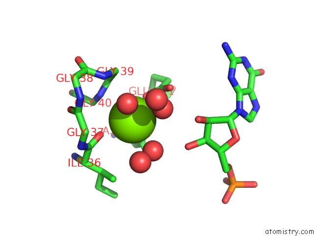
Mono view
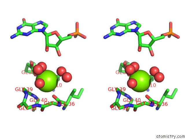
Stereo pair view
Mono view
Stereo pair view
A full contact list of Magnesium with other atoms in the Mg binding
site number 1 of Saccharomyces Cerevisiae Hypoxanthine-Guanine Phosphoribosyltransferase in Complex with Gmp (Monoclinic Crystal Form) within 5.0Å range:
|
Reference:
L.Moynie,
M.F.Giraud,
A.Breton,
F.Boissier,
B.Daignan-Fornier,
A.Dautant.
Functional Significance of Four Successive Glycine Residues in the Pyrophosphate Binding Loop of Fungal 6-Oxopurine Phosphoribosyltransferases. Protein Sci. V. 21 1185 2012.
ISSN: ISSN 0961-8368
PubMed: 22610485
DOI: 10.1002/PRO.2098
Page generated: Sun Aug 10 16:22:53 2025
ISSN: ISSN 0961-8368
PubMed: 22610485
DOI: 10.1002/PRO.2098
Last articles
Mg in 5ZKJMg in 5ZKI
Mg in 5ZK6
Mg in 5ZE9
Mg in 5ZFX
Mg in 5ZCT
Mg in 5ZE6
Mg in 5ZE4
Mg in 5ZDN
Mg in 5ZE0