Magnesium »
PDB 3dfy-3dts »
3dg6 »
Magnesium in PDB 3dg6: Crystal Structure of Muconate Lactonizing Enzyme From Mucobacterium Smegmatis Complexed with Muconolactone
Protein crystallography data
The structure of Crystal Structure of Muconate Lactonizing Enzyme From Mucobacterium Smegmatis Complexed with Muconolactone, PDB code: 3dg6
was solved by
A.A.Fedorov,
E.V.Fedorov,
A.Sakai,
J.A.Gerlt,
S.C.Almo,
with X-Ray Crystallography technique. A brief refinement statistics is given in the table below:
| Resolution Low / High (Å) | 24.38 / 1.60 |
| Space group | I 4 2 2 |
| Cell size a, b, c (Å), α, β, γ (°) | 123.840, 123.840, 117.370, 90.00, 90.00, 90.00 |
| R / Rfree (%) | 18.5 / 20.1 |
Magnesium Binding Sites:
The binding sites of Magnesium atom in the Crystal Structure of Muconate Lactonizing Enzyme From Mucobacterium Smegmatis Complexed with Muconolactone
(pdb code 3dg6). This binding sites where shown within
5.0 Angstroms radius around Magnesium atom.
In total only one binding site of Magnesium was determined in the Crystal Structure of Muconate Lactonizing Enzyme From Mucobacterium Smegmatis Complexed with Muconolactone, PDB code: 3dg6:
In total only one binding site of Magnesium was determined in the Crystal Structure of Muconate Lactonizing Enzyme From Mucobacterium Smegmatis Complexed with Muconolactone, PDB code: 3dg6:
Magnesium binding site 1 out of 1 in 3dg6
Go back to
Magnesium binding site 1 out
of 1 in the Crystal Structure of Muconate Lactonizing Enzyme From Mucobacterium Smegmatis Complexed with Muconolactone
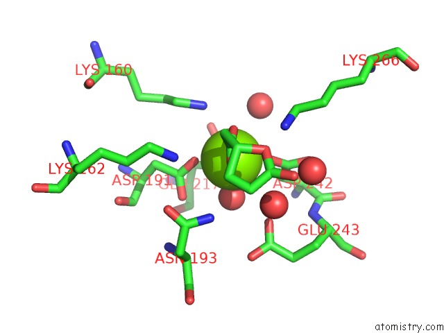
Mono view
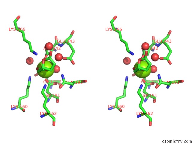
Stereo pair view
Mono view
Stereo pair view
A full contact list of Magnesium with other atoms in the Mg binding
site number 1 of Crystal Structure of Muconate Lactonizing Enzyme From Mucobacterium Smegmatis Complexed with Muconolactone within 5.0Å range:
|
Reference:
A.Sakai,
A.A.Fedorov,
E.V.Fedorov,
A.M.Schnoes,
M.E.Glasner,
S.Brown,
M.E.Rutter,
K.Bain,
S.Chang,
T.Gheyi,
J.M.Sauder,
S.K.Burley,
P.C.Babbitt,
S.C.Almo,
J.A.Gerlt.
Evolution of Enzymatic Activities in the Enolase Superfamily: Stereochemically Distinct Mechanisms in Two Families of Cis,Cis-Muconate Lactonizing Enzymes Biochemistry V. 48 1445 2009.
ISSN: ISSN 0006-2960
PubMed: 19220063
DOI: 10.1021/BI802277H
Page generated: Sun Aug 10 20:05:58 2025
ISSN: ISSN 0006-2960
PubMed: 19220063
DOI: 10.1021/BI802277H
Last articles
Mg in 5Y85Mg in 5Y9Z
Mg in 5Y8V
Mg in 5Y88
Mg in 5Y8H
Mg in 5Y8B
Mg in 5Y5P
Mg in 5Y5Q
Mg in 5Y6Z
Mg in 5XYM