Magnesium »
PDB 3m1v-3mfh »
3mco »
Magnesium in PDB 3mco: Crystal Structure of the 6-Hyroxymethyl-7,8-Dihydropterin Pyrophosphokinase Dihydropteroate Synthase Bifunctional Enzyme From Francisella Tularensis
Protein crystallography data
The structure of Crystal Structure of the 6-Hyroxymethyl-7,8-Dihydropterin Pyrophosphokinase Dihydropteroate Synthase Bifunctional Enzyme From Francisella Tularensis, PDB code: 3mco
was solved by
C.W.Pemble Iv,
P.K.Mehta,
S.Mehra,
Z.Li,
R.E.Lee,
S.W.White,
with X-Ray Crystallography technique. A brief refinement statistics is given in the table below:
| Resolution Low / High (Å) | 38.25 / 2.30 |
| Space group | P 1 |
| Cell size a, b, c (Å), α, β, γ (°) | 42.521, 58.472, 109.327, 82.00, 81.00, 68.08 |
| R / Rfree (%) | 24.1 / 29.4 |
Magnesium Binding Sites:
The binding sites of Magnesium atom in the Crystal Structure of the 6-Hyroxymethyl-7,8-Dihydropterin Pyrophosphokinase Dihydropteroate Synthase Bifunctional Enzyme From Francisella Tularensis
(pdb code 3mco). This binding sites where shown within
5.0 Angstroms radius around Magnesium atom.
In total 4 binding sites of Magnesium where determined in the Crystal Structure of the 6-Hyroxymethyl-7,8-Dihydropterin Pyrophosphokinase Dihydropteroate Synthase Bifunctional Enzyme From Francisella Tularensis, PDB code: 3mco:
Jump to Magnesium binding site number: 1; 2; 3; 4;
In total 4 binding sites of Magnesium where determined in the Crystal Structure of the 6-Hyroxymethyl-7,8-Dihydropterin Pyrophosphokinase Dihydropteroate Synthase Bifunctional Enzyme From Francisella Tularensis, PDB code: 3mco:
Jump to Magnesium binding site number: 1; 2; 3; 4;
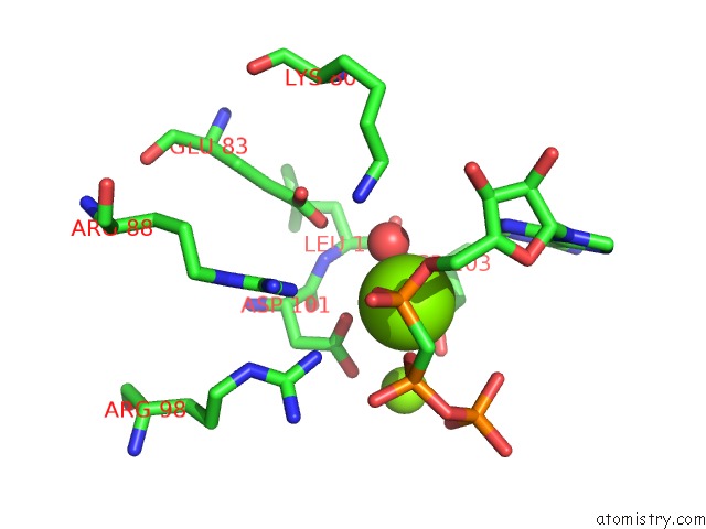
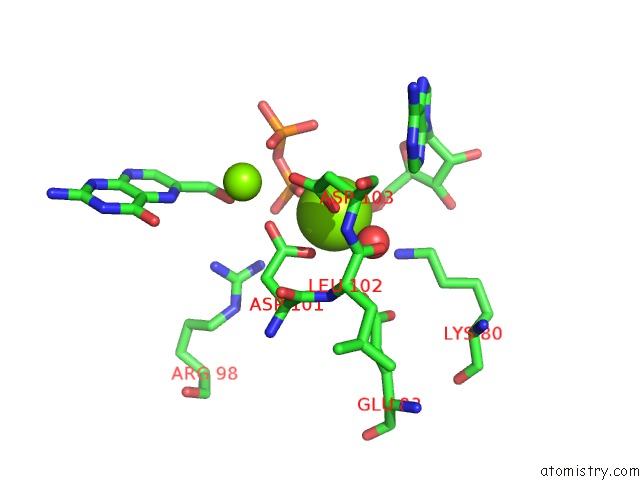
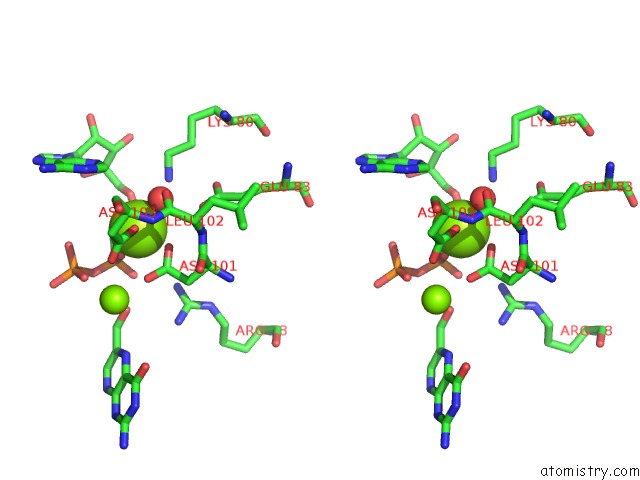
Magnesium binding site 1 out of 4 in 3mco
Go back to
Magnesium binding site 1 out
of 4 in the Crystal Structure of the 6-Hyroxymethyl-7,8-Dihydropterin Pyrophosphokinase Dihydropteroate Synthase Bifunctional Enzyme From Francisella Tularensis
Mono view
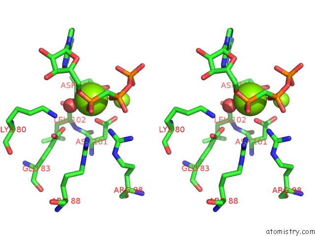
Stereo pair view
Mono view
Stereo pair view
A full contact list of Magnesium with other atoms in the Mg binding
site number 1 of Crystal Structure of the 6-Hyroxymethyl-7,8-Dihydropterin Pyrophosphokinase Dihydropteroate Synthase Bifunctional Enzyme From Francisella Tularensis within 5.0Å range:
|
Magnesium binding site 2 out of 4 in 3mco
Go back to
Magnesium binding site 2 out
of 4 in the Crystal Structure of the 6-Hyroxymethyl-7,8-Dihydropterin Pyrophosphokinase Dihydropteroate Synthase Bifunctional Enzyme From Francisella Tularensis
Mono view
Stereo pair view
Mono view
Stereo pair view
A full contact list of Magnesium with other atoms in the Mg binding
site number 2 of Crystal Structure of the 6-Hyroxymethyl-7,8-Dihydropterin Pyrophosphokinase Dihydropteroate Synthase Bifunctional Enzyme From Francisella Tularensis within 5.0Å range:
|
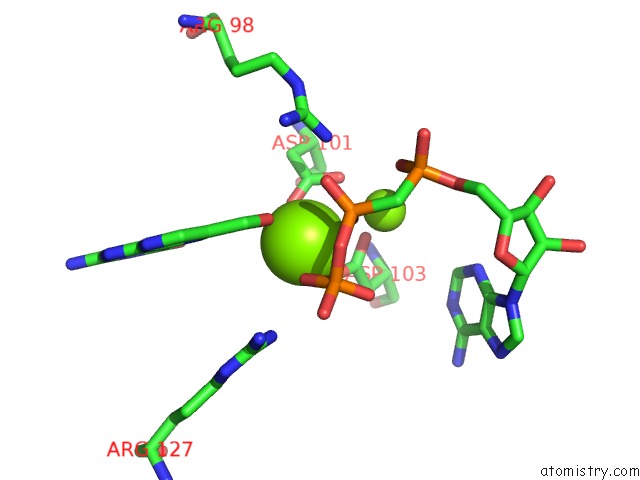
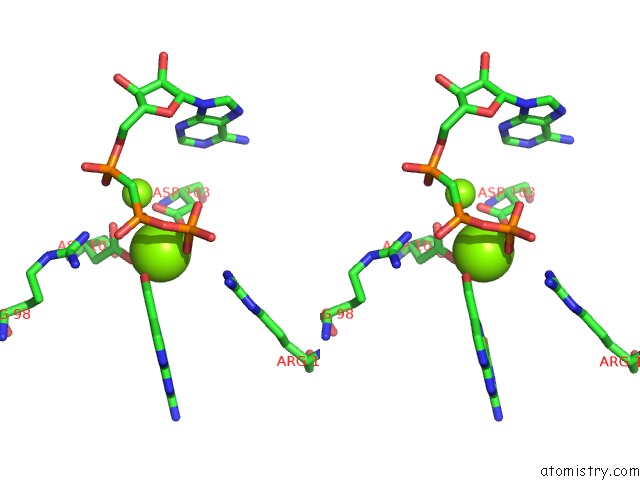
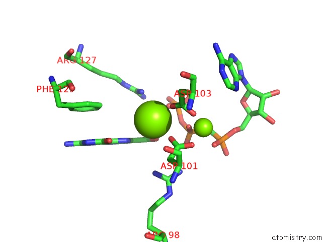
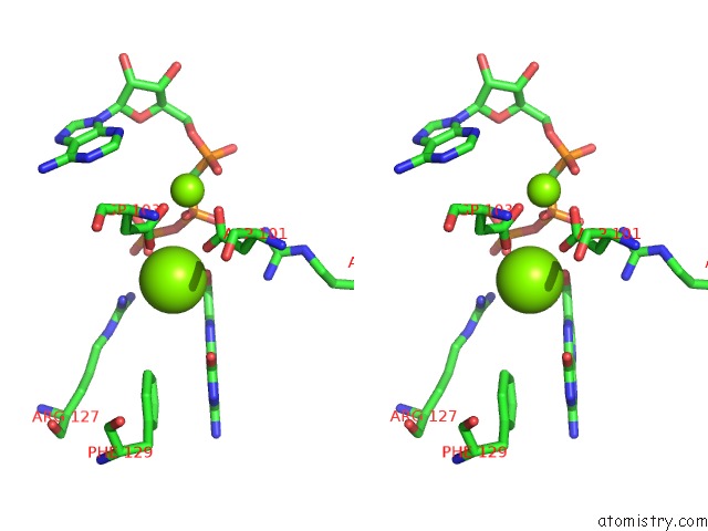
Magnesium binding site 3 out of 4 in 3mco
Go back to
Magnesium binding site 3 out
of 4 in the Crystal Structure of the 6-Hyroxymethyl-7,8-Dihydropterin Pyrophosphokinase Dihydropteroate Synthase Bifunctional Enzyme From Francisella Tularensis
Mono view
Stereo pair view
Mono view
Stereo pair view
A full contact list of Magnesium with other atoms in the Mg binding
site number 3 of Crystal Structure of the 6-Hyroxymethyl-7,8-Dihydropterin Pyrophosphokinase Dihydropteroate Synthase Bifunctional Enzyme From Francisella Tularensis within 5.0Å range:
|
Magnesium binding site 4 out of 4 in 3mco
Go back to
Magnesium binding site 4 out
of 4 in the Crystal Structure of the 6-Hyroxymethyl-7,8-Dihydropterin Pyrophosphokinase Dihydropteroate Synthase Bifunctional Enzyme From Francisella Tularensis
Mono view
Stereo pair view
Mono view
Stereo pair view
A full contact list of Magnesium with other atoms in the Mg binding
site number 4 of Crystal Structure of the 6-Hyroxymethyl-7,8-Dihydropterin Pyrophosphokinase Dihydropteroate Synthase Bifunctional Enzyme From Francisella Tularensis within 5.0Å range:
|
Reference:
C.W.Pemble,
P.K.Mehta,
S.Mehra,
Z.Li,
A.Nourse,
R.E.Lee,
S.W.White.
Crystal Structure of the 6-Hydroxymethyl-7,8-Dihydropterin Pyrophosphokinase.Dihydropteroate Synthase Bifunctional Enzyme From Francisella Tularensis. Plos One V. 5 14165 2010.
ISSN: ESSN 1932-6203
PubMed: 21152407
DOI: 10.1371/JOURNAL.PONE.0014165
Page generated: Mon Aug 11 00:31:26 2025
ISSN: ESSN 1932-6203
PubMed: 21152407
DOI: 10.1371/JOURNAL.PONE.0014165
Last articles
Mg in 5YUTMg in 5YUS
Mg in 5YU8
Mg in 5YU6
Mg in 5YTY
Mg in 5YTG
Mg in 5YTH
Mg in 5YTF
Mg in 5YTD
Mg in 5YTE