Magnesium »
PDB 3mv1-3n8b »
3n0s »
Magnesium in PDB 3n0s: Crystal Structure of BA2930 Mutant (H183A) in Complex with Accoa
Enzymatic activity of Crystal Structure of BA2930 Mutant (H183A) in Complex with Accoa
All present enzymatic activity of Crystal Structure of BA2930 Mutant (H183A) in Complex with Accoa:
2.3.1.81;
2.3.1.81;
Protein crystallography data
The structure of Crystal Structure of BA2930 Mutant (H183A) in Complex with Accoa, PDB code: 3n0s
was solved by
M.M.Klimecka,
M.Chruszcz,
P.J.Porebski,
M.Cymborowski,
W.F.Anderson,
W.Minor,
with X-Ray Crystallography technique. A brief refinement statistics is given in the table below:
| Resolution Low / High (Å) | 50.00 / 2.15 |
| Space group | P 1 21 1 |
| Cell size a, b, c (Å), α, β, γ (°) | 72.036, 109.441, 74.048, 90.00, 111.86, 90.00 |
| R / Rfree (%) | 17.3 / 22.8 |
Magnesium Binding Sites:
The binding sites of Magnesium atom in the Crystal Structure of BA2930 Mutant (H183A) in Complex with Accoa
(pdb code 3n0s). This binding sites where shown within
5.0 Angstroms radius around Magnesium atom.
In total 2 binding sites of Magnesium where determined in the Crystal Structure of BA2930 Mutant (H183A) in Complex with Accoa, PDB code: 3n0s:
Jump to Magnesium binding site number: 1; 2;
In total 2 binding sites of Magnesium where determined in the Crystal Structure of BA2930 Mutant (H183A) in Complex with Accoa, PDB code: 3n0s:
Jump to Magnesium binding site number: 1; 2;
Magnesium binding site 1 out of 2 in 3n0s
Go back to
Magnesium binding site 1 out
of 2 in the Crystal Structure of BA2930 Mutant (H183A) in Complex with Accoa
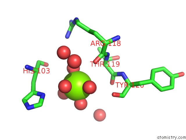
Mono view
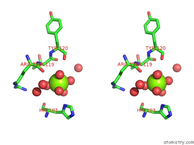
Stereo pair view
Mono view
Stereo pair view
A full contact list of Magnesium with other atoms in the Mg binding
site number 1 of Crystal Structure of BA2930 Mutant (H183A) in Complex with Accoa within 5.0Å range:
|
Magnesium binding site 2 out of 2 in 3n0s
Go back to
Magnesium binding site 2 out
of 2 in the Crystal Structure of BA2930 Mutant (H183A) in Complex with Accoa
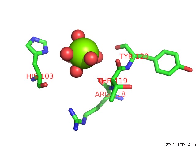
Mono view
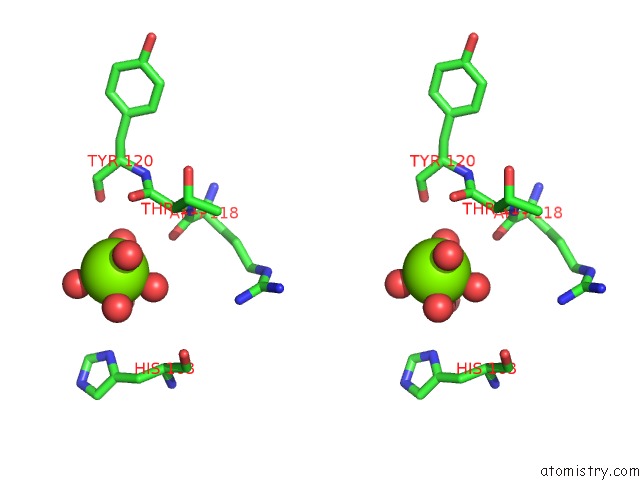
Stereo pair view
Mono view
Stereo pair view
A full contact list of Magnesium with other atoms in the Mg binding
site number 2 of Crystal Structure of BA2930 Mutant (H183A) in Complex with Accoa within 5.0Å range:
|
Reference:
M.M.Klimecka,
M.Chruszcz,
J.Font,
T.Skarina,
I.Shumilin,
O.Onopryienko,
P.J.Porebski,
M.Cymborowski,
M.D.Zimmerman,
J.Hasseman,
I.J.Glomski,
L.Lebioda,
A.Savchenko,
A.Edwards,
W.Minor.
Structural Analysis of A Putative Aminoglycoside N-Acetyltransferase From Bacillus Anthracis. J.Mol.Biol. V. 410 411 2011.
ISSN: ISSN 0022-2836
PubMed: 21601576
DOI: 10.1016/J.JMB.2011.04.076
Page generated: Mon Aug 11 00:45:45 2025
ISSN: ISSN 0022-2836
PubMed: 21601576
DOI: 10.1016/J.JMB.2011.04.076
Last articles
Mg in 5ZKJMg in 5ZKI
Mg in 5ZK6
Mg in 5ZE9
Mg in 5ZFX
Mg in 5ZCT
Mg in 5ZE6
Mg in 5ZE4
Mg in 5ZDN
Mg in 5ZE0