Magnesium »
PDB 3py8-3q86 »
3q45 »
Magnesium in PDB 3q45: Crystal Structure of Dipeptide Epimerase From Cytophaga Hutchinsonii Complexed with Mg and Dipeptide D-Ala-L-Val
Enzymatic activity of Crystal Structure of Dipeptide Epimerase From Cytophaga Hutchinsonii Complexed with Mg and Dipeptide D-Ala-L-Val
All present enzymatic activity of Crystal Structure of Dipeptide Epimerase From Cytophaga Hutchinsonii Complexed with Mg and Dipeptide D-Ala-L-Val:
5.5.1.7;
5.5.1.7;
Protein crystallography data
The structure of Crystal Structure of Dipeptide Epimerase From Cytophaga Hutchinsonii Complexed with Mg and Dipeptide D-Ala-L-Val, PDB code: 3q45
was solved by
T.Lukk,
J.A.Gerlt,
S.K.Nair,
with X-Ray Crystallography technique. A brief refinement statistics is given in the table below:
| Resolution Low / High (Å) | 20.00 / 3.00 |
| Space group | P 1 21 1 |
| Cell size a, b, c (Å), α, β, γ (°) | 94.000, 158.200, 182.740, 90.00, 100.28, 90.00 |
| R / Rfree (%) | 22.7 / 25.1 |
Magnesium Binding Sites:
The binding sites of Magnesium atom in the Crystal Structure of Dipeptide Epimerase From Cytophaga Hutchinsonii Complexed with Mg and Dipeptide D-Ala-L-Val
(pdb code 3q45). This binding sites where shown within
5.0 Angstroms radius around Magnesium atom.
In total 9 binding sites of Magnesium where determined in the Crystal Structure of Dipeptide Epimerase From Cytophaga Hutchinsonii Complexed with Mg and Dipeptide D-Ala-L-Val, PDB code: 3q45:
Jump to Magnesium binding site number: 1; 2; 3; 4; 5; 6; 7; 8; 9;
In total 9 binding sites of Magnesium where determined in the Crystal Structure of Dipeptide Epimerase From Cytophaga Hutchinsonii Complexed with Mg and Dipeptide D-Ala-L-Val, PDB code: 3q45:
Jump to Magnesium binding site number: 1; 2; 3; 4; 5; 6; 7; 8; 9;
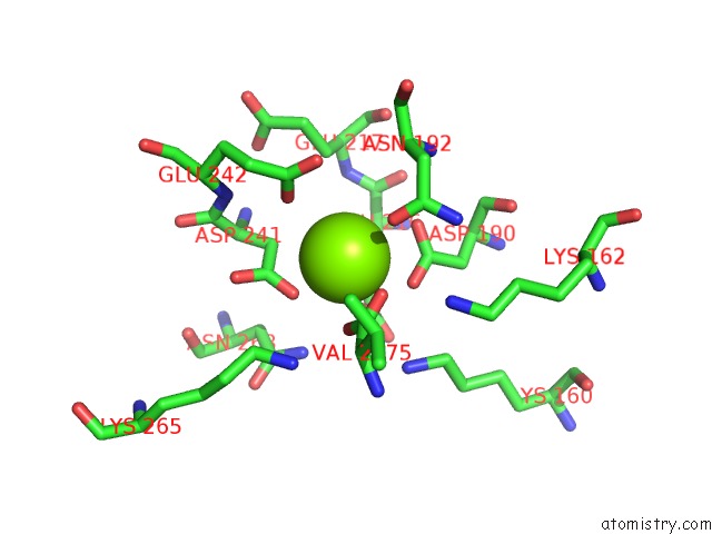
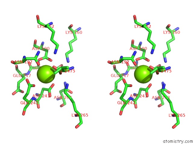
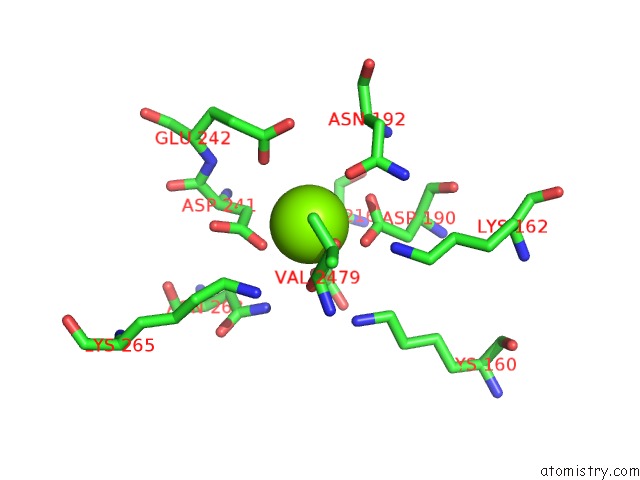
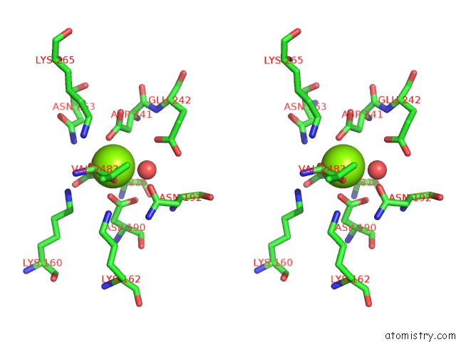
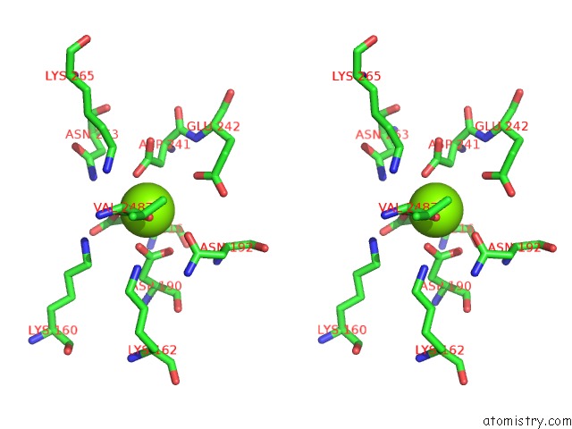
Magnesium binding site 1 out of 9 in 3q45
Go back to
Magnesium binding site 1 out
of 9 in the Crystal Structure of Dipeptide Epimerase From Cytophaga Hutchinsonii Complexed with Mg and Dipeptide D-Ala-L-Val
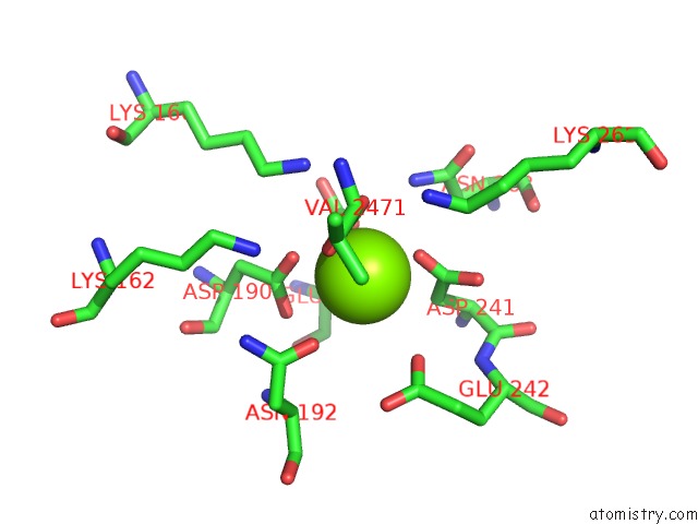
Mono view
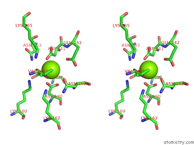
Stereo pair view
Mono view
Stereo pair view
A full contact list of Magnesium with other atoms in the Mg binding
site number 1 of Crystal Structure of Dipeptide Epimerase From Cytophaga Hutchinsonii Complexed with Mg and Dipeptide D-Ala-L-Val within 5.0Å range:
|
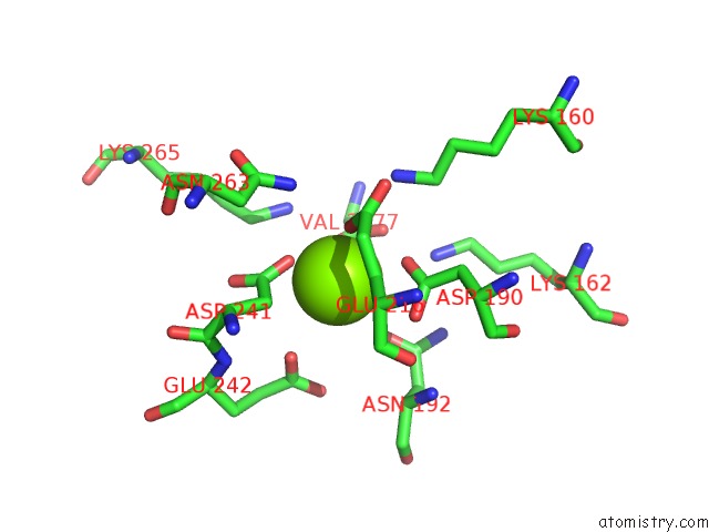
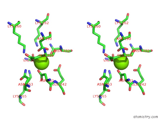
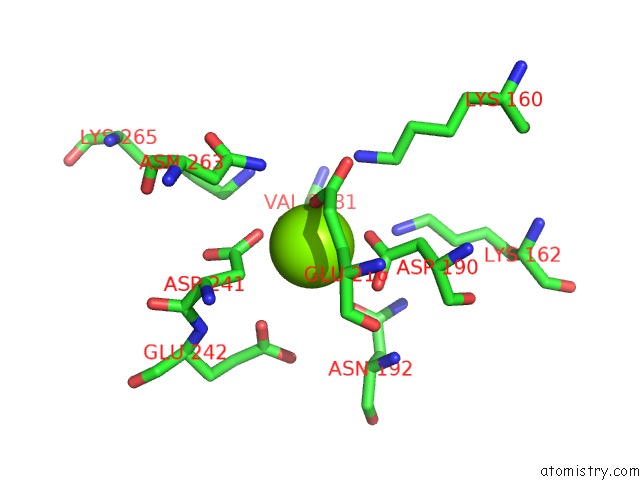
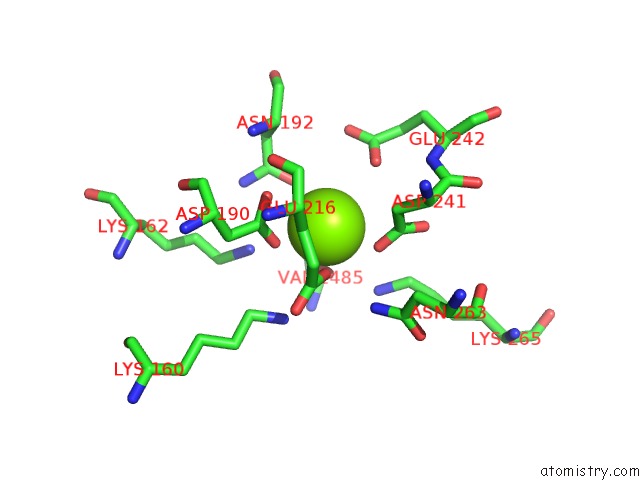
Magnesium binding site 2 out of 9 in 3q45
Go back to
Magnesium binding site 2 out
of 9 in the Crystal Structure of Dipeptide Epimerase From Cytophaga Hutchinsonii Complexed with Mg and Dipeptide D-Ala-L-Val
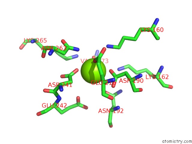
Mono view
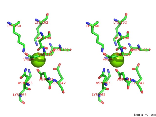
Stereo pair view
Mono view
Stereo pair view
A full contact list of Magnesium with other atoms in the Mg binding
site number 2 of Crystal Structure of Dipeptide Epimerase From Cytophaga Hutchinsonii Complexed with Mg and Dipeptide D-Ala-L-Val within 5.0Å range:
|
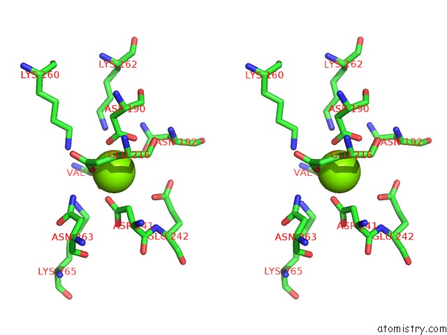
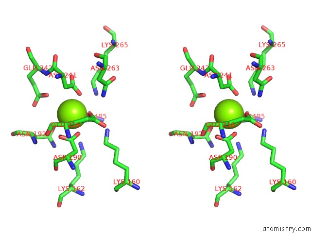
Magnesium binding site 3 out of 9 in 3q45
Go back to
Magnesium binding site 3 out
of 9 in the Crystal Structure of Dipeptide Epimerase From Cytophaga Hutchinsonii Complexed with Mg and Dipeptide D-Ala-L-Val
Mono view
Stereo pair view
Mono view
Stereo pair view
A full contact list of Magnesium with other atoms in the Mg binding
site number 3 of Crystal Structure of Dipeptide Epimerase From Cytophaga Hutchinsonii Complexed with Mg and Dipeptide D-Ala-L-Val within 5.0Å range:
|
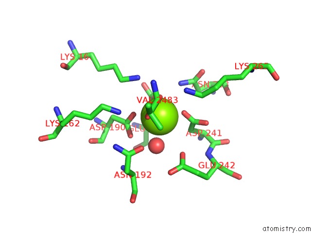
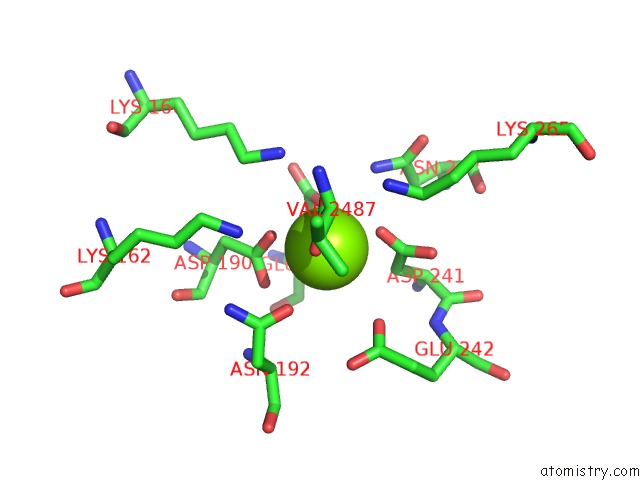
Magnesium binding site 4 out of 9 in 3q45
Go back to
Magnesium binding site 4 out
of 9 in the Crystal Structure of Dipeptide Epimerase From Cytophaga Hutchinsonii Complexed with Mg and Dipeptide D-Ala-L-Val
Mono view
Stereo pair view
Mono view
Stereo pair view
A full contact list of Magnesium with other atoms in the Mg binding
site number 4 of Crystal Structure of Dipeptide Epimerase From Cytophaga Hutchinsonii Complexed with Mg and Dipeptide D-Ala-L-Val within 5.0Å range:
|
Magnesium binding site 5 out of 9 in 3q45
Go back to
Magnesium binding site 5 out
of 9 in the Crystal Structure of Dipeptide Epimerase From Cytophaga Hutchinsonii Complexed with Mg and Dipeptide D-Ala-L-Val
Mono view

Stereo pair view
Mono view
Stereo pair view
A full contact list of Magnesium with other atoms in the Mg binding
site number 5 of Crystal Structure of Dipeptide Epimerase From Cytophaga Hutchinsonii Complexed with Mg and Dipeptide D-Ala-L-Val within 5.0Å range:
|
Magnesium binding site 6 out of 9 in 3q45
Go back to
Magnesium binding site 6 out
of 9 in the Crystal Structure of Dipeptide Epimerase From Cytophaga Hutchinsonii Complexed with Mg and Dipeptide D-Ala-L-Val
Mono view
Stereo pair view
Mono view
Stereo pair view
A full contact list of Magnesium with other atoms in the Mg binding
site number 6 of Crystal Structure of Dipeptide Epimerase From Cytophaga Hutchinsonii Complexed with Mg and Dipeptide D-Ala-L-Val within 5.0Å range:
|
Magnesium binding site 7 out of 9 in 3q45
Go back to
Magnesium binding site 7 out
of 9 in the Crystal Structure of Dipeptide Epimerase From Cytophaga Hutchinsonii Complexed with Mg and Dipeptide D-Ala-L-Val
Mono view
Stereo pair view
Mono view
Stereo pair view
A full contact list of Magnesium with other atoms in the Mg binding
site number 7 of Crystal Structure of Dipeptide Epimerase From Cytophaga Hutchinsonii Complexed with Mg and Dipeptide D-Ala-L-Val within 5.0Å range:
|
Magnesium binding site 8 out of 9 in 3q45
Go back to
Magnesium binding site 8 out
of 9 in the Crystal Structure of Dipeptide Epimerase From Cytophaga Hutchinsonii Complexed with Mg and Dipeptide D-Ala-L-Val
Mono view
Stereo pair view
Mono view
Stereo pair view
A full contact list of Magnesium with other atoms in the Mg binding
site number 8 of Crystal Structure of Dipeptide Epimerase From Cytophaga Hutchinsonii Complexed with Mg and Dipeptide D-Ala-L-Val within 5.0Å range:
|
Magnesium binding site 9 out of 9 in 3q45
Go back to
Magnesium binding site 9 out
of 9 in the Crystal Structure of Dipeptide Epimerase From Cytophaga Hutchinsonii Complexed with Mg and Dipeptide D-Ala-L-Val
Mono view
Stereo pair view
Mono view
Stereo pair view
A full contact list of Magnesium with other atoms in the Mg binding
site number 9 of Crystal Structure of Dipeptide Epimerase From Cytophaga Hutchinsonii Complexed with Mg and Dipeptide D-Ala-L-Val within 5.0Å range:
|
Reference:
T.Lukk,
A.Sakai,
C.Kalyanaraman,
S.D.Brown,
H.J.Imker,
L.Song,
A.A.Fedorov,
E.V.Fedorov,
R.Toro,
B.Hillerich,
R.Seidel,
Y.Patskovsky,
M.W.Vetting,
S.K.Nair,
P.C.Babbitt,
S.C.Almo,
J.A.Gerlt,
M.P.Jacobson.
Homology Models Guide Discovery of Diverse Enzyme Specificities Among Dipeptide Epimerases in the Enolase Superfamily. Proc.Natl.Acad.Sci.Usa V. 109 4122 2012.
ISSN: ISSN 0027-8424
PubMed: 22392983
DOI: 10.1073/PNAS.1112081109
Page generated: Mon Aug 11 02:17:42 2025
ISSN: ISSN 0027-8424
PubMed: 22392983
DOI: 10.1073/PNAS.1112081109
Last articles
Mg in 4JHDMg in 4JH6
Mg in 4JH8
Mg in 4JH7
Mg in 4JH3
Mg in 4JH5
Mg in 4JF2
Mg in 4JH2
Mg in 4JH1
Mg in 4JEJ