Magnesium »
PDB 3rer-3rr5 »
3rkq »
Magnesium in PDB 3rkq: NKX2.5 Homeodomain Dimer Bound to Anf-242 Dna
Protein crystallography data
The structure of NKX2.5 Homeodomain Dimer Bound to Anf-242 Dna, PDB code: 3rkq
was solved by
C.Genis,
P.Scone,
H.Kasahara,
H.-J.Nam,
with X-Ray Crystallography technique. A brief refinement statistics is given in the table below:
| Resolution Low / High (Å) | 30.94 / 1.70 |
| Space group | P 65 |
| Cell size a, b, c (Å), α, β, γ (°) | 71.450, 71.450, 94.330, 90.00, 90.00, 120.00 |
| R / Rfree (%) | 21.1 / 25.7 |
Magnesium Binding Sites:
The binding sites of Magnesium atom in the NKX2.5 Homeodomain Dimer Bound to Anf-242 Dna
(pdb code 3rkq). This binding sites where shown within
5.0 Angstroms radius around Magnesium atom.
In total only one binding site of Magnesium was determined in the NKX2.5 Homeodomain Dimer Bound to Anf-242 Dna, PDB code: 3rkq:
In total only one binding site of Magnesium was determined in the NKX2.5 Homeodomain Dimer Bound to Anf-242 Dna, PDB code: 3rkq:
Magnesium binding site 1 out of 1 in 3rkq
Go back to
Magnesium binding site 1 out
of 1 in the NKX2.5 Homeodomain Dimer Bound to Anf-242 Dna
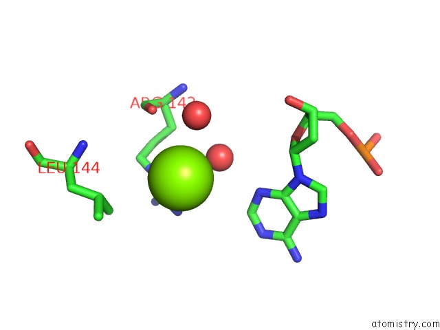
Mono view
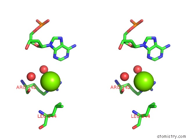
Stereo pair view
Mono view
Stereo pair view
A full contact list of Magnesium with other atoms in the Mg binding
site number 1 of NKX2.5 Homeodomain Dimer Bound to Anf-242 Dna within 5.0Å range:
|
Reference:
L.Pradhan,
C.Genis,
P.Scone,
E.O.Weinberg,
H.Kasahara,
H.J.Nam.
Crystal Structure of the Human NKX2.5 Homeodomain in Complex with Dna Target. Biochemistry V. 51 6312 2012.
ISSN: ISSN 0006-2960
PubMed: 22849347
DOI: 10.1021/BI300849C
Page generated: Mon Aug 11 02:45:24 2025
ISSN: ISSN 0006-2960
PubMed: 22849347
DOI: 10.1021/BI300849C
Last articles
Mg in 6OEMMg in 6OEW
Mg in 6OD9
Mg in 6OBJ
Mg in 6OE3
Mg in 6OB3
Mg in 6OAP
Mg in 6OAQ
Mg in 6OB2
Mg in 6O9P