Magnesium »
PDB 3umy-3v4i »
3uqe »
Magnesium in PDB 3uqe: Crystal Structure of the Phosphofructokinase-2 Mutant Y23D From Escherichia Coli
Enzymatic activity of Crystal Structure of the Phosphofructokinase-2 Mutant Y23D From Escherichia Coli
All present enzymatic activity of Crystal Structure of the Phosphofructokinase-2 Mutant Y23D From Escherichia Coli:
2.7.1.11;
2.7.1.11;
Protein crystallography data
The structure of Crystal Structure of the Phosphofructokinase-2 Mutant Y23D From Escherichia Coli, PDB code: 3uqe
was solved by
H.M.Pereira,
A.Caniuguir,
M.Baez,
R.Cabrera,
R.C.Garatt,
J.Babul,
with X-Ray Crystallography technique. A brief refinement statistics is given in the table below:
| Resolution Low / High (Å) | 48.98 / 2.20 |
| Space group | P 2 2 21 |
| Cell size a, b, c (Å), α, β, γ (°) | 43.838, 89.123, 175.908, 90.00, 90.00, 90.00 |
| R / Rfree (%) | 21.9 / 25.3 |
Magnesium Binding Sites:
The binding sites of Magnesium atom in the Crystal Structure of the Phosphofructokinase-2 Mutant Y23D From Escherichia Coli
(pdb code 3uqe). This binding sites where shown within
5.0 Angstroms radius around Magnesium atom.
In total 4 binding sites of Magnesium where determined in the Crystal Structure of the Phosphofructokinase-2 Mutant Y23D From Escherichia Coli, PDB code: 3uqe:
Jump to Magnesium binding site number: 1; 2; 3; 4;
In total 4 binding sites of Magnesium where determined in the Crystal Structure of the Phosphofructokinase-2 Mutant Y23D From Escherichia Coli, PDB code: 3uqe:
Jump to Magnesium binding site number: 1; 2; 3; 4;
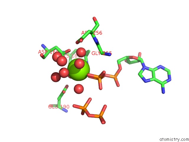
Magnesium binding site 1 out of 4 in 3uqe
Go back to
Magnesium binding site 1 out
of 4 in the Crystal Structure of the Phosphofructokinase-2 Mutant Y23D From Escherichia Coli
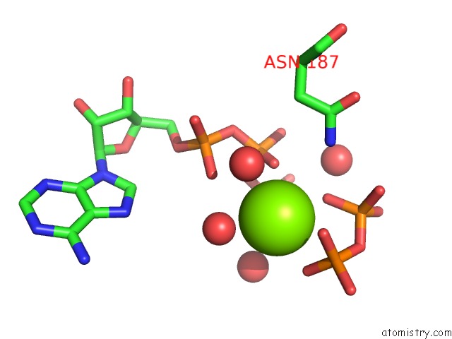
Mono view
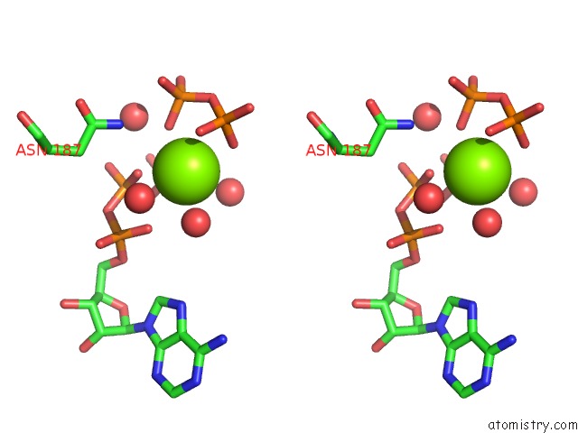
Stereo pair view
Mono view
Stereo pair view
A full contact list of Magnesium with other atoms in the Mg binding
site number 1 of Crystal Structure of the Phosphofructokinase-2 Mutant Y23D From Escherichia Coli within 5.0Å range:
|
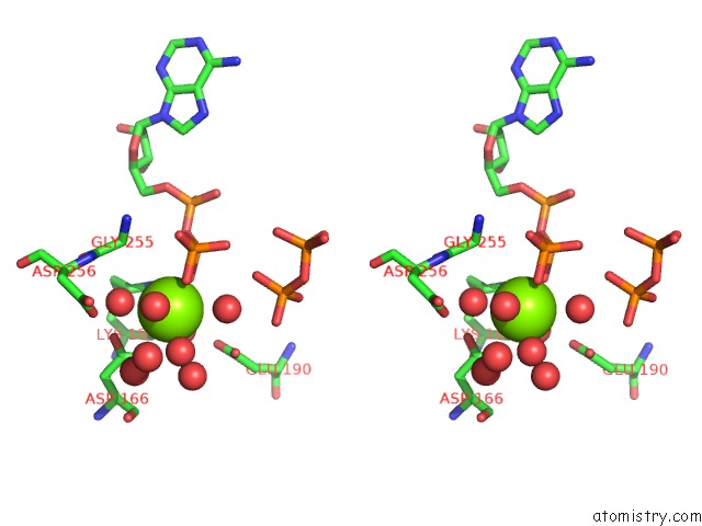
Magnesium binding site 2 out of 4 in 3uqe
Go back to
Magnesium binding site 2 out
of 4 in the Crystal Structure of the Phosphofructokinase-2 Mutant Y23D From Escherichia Coli
Mono view
Stereo pair view
Mono view
Stereo pair view
A full contact list of Magnesium with other atoms in the Mg binding
site number 2 of Crystal Structure of the Phosphofructokinase-2 Mutant Y23D From Escherichia Coli within 5.0Å range:
|
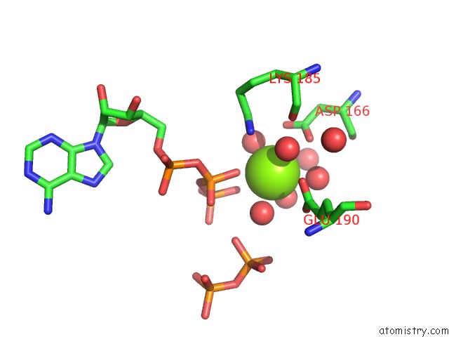
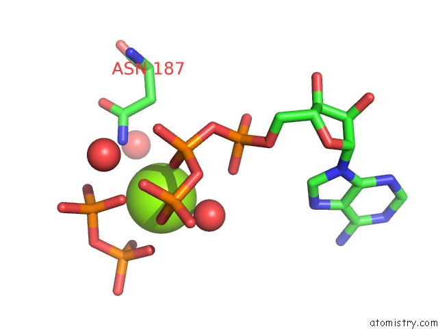
Magnesium binding site 3 out of 4 in 3uqe
Go back to
Magnesium binding site 3 out
of 4 in the Crystal Structure of the Phosphofructokinase-2 Mutant Y23D From Escherichia Coli
Mono view
Stereo pair view
Mono view
Stereo pair view
A full contact list of Magnesium with other atoms in the Mg binding
site number 3 of Crystal Structure of the Phosphofructokinase-2 Mutant Y23D From Escherichia Coli within 5.0Å range:
|
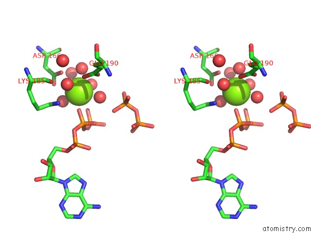
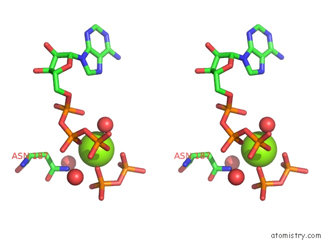
Magnesium binding site 4 out of 4 in 3uqe
Go back to
Magnesium binding site 4 out
of 4 in the Crystal Structure of the Phosphofructokinase-2 Mutant Y23D From Escherichia Coli
Mono view
Stereo pair view
Mono view
Stereo pair view
A full contact list of Magnesium with other atoms in the Mg binding
site number 4 of Crystal Structure of the Phosphofructokinase-2 Mutant Y23D From Escherichia Coli within 5.0Å range:
|
Reference:
H.M.Pereira,
A.Caniuguir,
M.Baez,
R.Cabrera,
R.C.Garratt,
J.Babul.
Structure of E. Coli PFK2 Mutant Y23D To Be Published.
Page generated: Mon Aug 11 04:18:40 2025
Last articles
Mg in 4Q39Mg in 4Q2G
Mg in 4Q2E
Mg in 4Q23
Mg in 4Q15
Mg in 4Q1V
Mg in 4Q2D
Mg in 4Q21
Mg in 4Q01
Mg in 4Q04