Magnesium »
PDB 4c30-4cgk »
4c30 »
Magnesium in PDB 4c30: Crystal Structure of Deinococcus Radiodurans Uvrd in Complex with Dna, Form 2
Enzymatic activity of Crystal Structure of Deinococcus Radiodurans Uvrd in Complex with Dna, Form 2
All present enzymatic activity of Crystal Structure of Deinococcus Radiodurans Uvrd in Complex with Dna, Form 2:
3.6.4.12;
3.6.4.12;
Protein crystallography data
The structure of Crystal Structure of Deinococcus Radiodurans Uvrd in Complex with Dna, Form 2, PDB code: 4c30
was solved by
M.Stelter,
S.Acajjaoui,
S.Mcsweeney,
J.Timmins,
with X-Ray Crystallography technique. A brief refinement statistics is given in the table below:
| Resolution Low / High (Å) | 66.18 / 3.00 |
| Space group | P 1 21 1 |
| Cell size a, b, c (Å), α, β, γ (°) | 68.489, 89.785, 293.798, 90.00, 89.97, 90.00 |
| R / Rfree (%) | 22.82 / 28.783 |
Magnesium Binding Sites:
The binding sites of Magnesium atom in the Crystal Structure of Deinococcus Radiodurans Uvrd in Complex with Dna, Form 2
(pdb code 4c30). This binding sites where shown within
5.0 Angstroms radius around Magnesium atom.
In total 6 binding sites of Magnesium where determined in the Crystal Structure of Deinococcus Radiodurans Uvrd in Complex with Dna, Form 2, PDB code: 4c30:
Jump to Magnesium binding site number: 1; 2; 3; 4; 5; 6;
In total 6 binding sites of Magnesium where determined in the Crystal Structure of Deinococcus Radiodurans Uvrd in Complex with Dna, Form 2, PDB code: 4c30:
Jump to Magnesium binding site number: 1; 2; 3; 4; 5; 6;
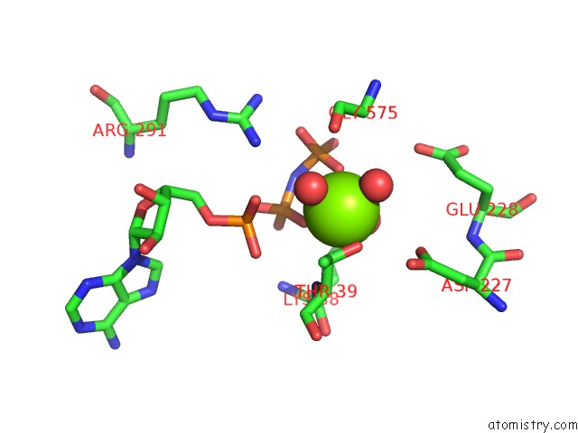
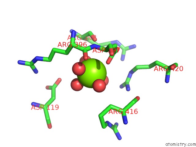
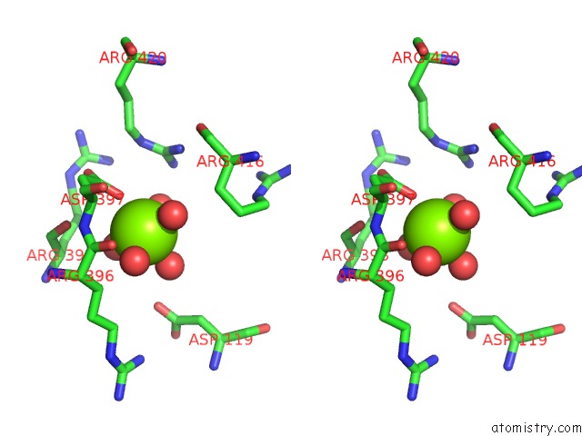
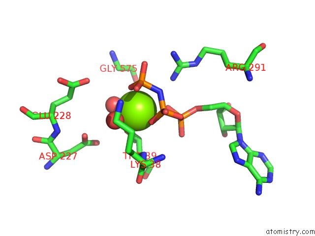
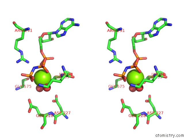
Magnesium binding site 1 out of 6 in 4c30
Go back to
Magnesium binding site 1 out
of 6 in the Crystal Structure of Deinococcus Radiodurans Uvrd in Complex with Dna, Form 2
Mono view
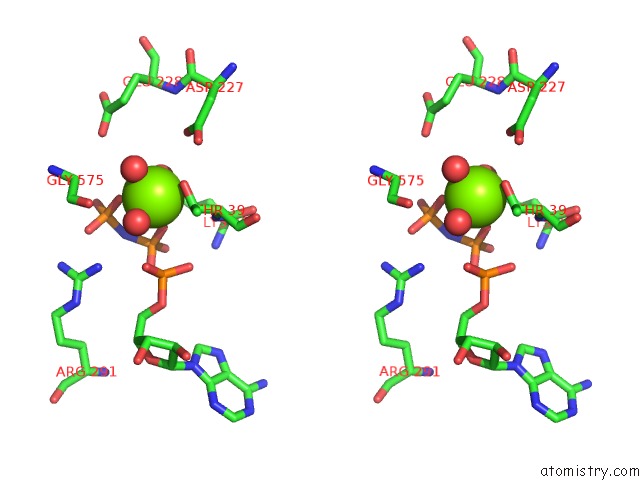
Stereo pair view
Mono view
Stereo pair view
A full contact list of Magnesium with other atoms in the Mg binding
site number 1 of Crystal Structure of Deinococcus Radiodurans Uvrd in Complex with Dna, Form 2 within 5.0Å range:
|
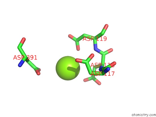
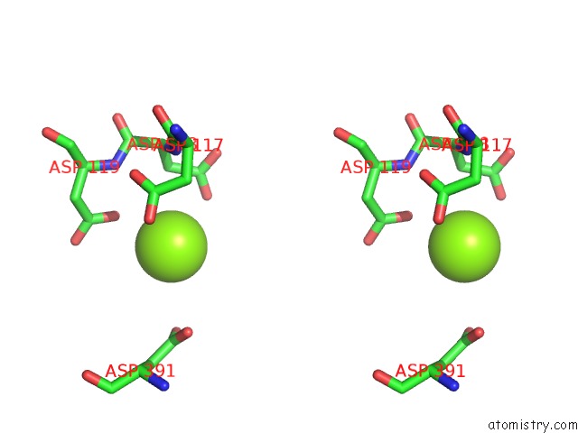
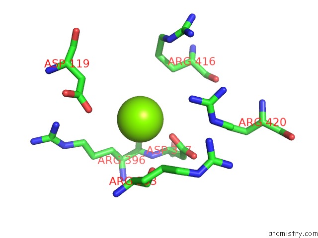
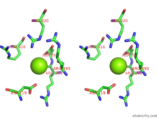
Magnesium binding site 2 out of 6 in 4c30
Go back to
Magnesium binding site 2 out
of 6 in the Crystal Structure of Deinococcus Radiodurans Uvrd in Complex with Dna, Form 2
Mono view
Stereo pair view
Mono view
Stereo pair view
A full contact list of Magnesium with other atoms in the Mg binding
site number 2 of Crystal Structure of Deinococcus Radiodurans Uvrd in Complex with Dna, Form 2 within 5.0Å range:
|
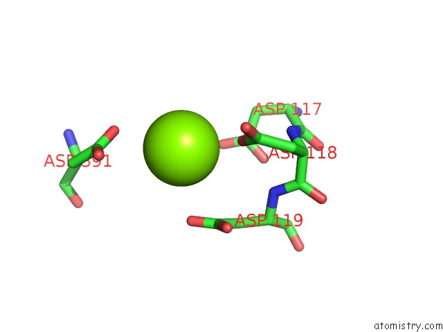
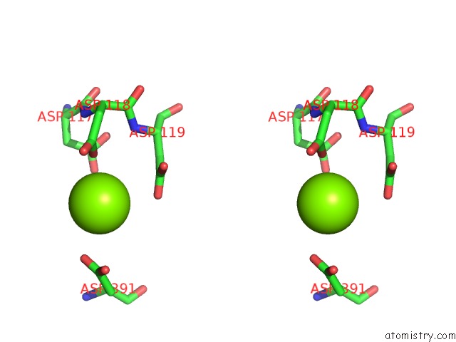
Magnesium binding site 3 out of 6 in 4c30
Go back to
Magnesium binding site 3 out
of 6 in the Crystal Structure of Deinococcus Radiodurans Uvrd in Complex with Dna, Form 2
Mono view
Stereo pair view
Mono view
Stereo pair view
A full contact list of Magnesium with other atoms in the Mg binding
site number 3 of Crystal Structure of Deinococcus Radiodurans Uvrd in Complex with Dna, Form 2 within 5.0Å range:
|
Magnesium binding site 4 out of 6 in 4c30
Go back to
Magnesium binding site 4 out
of 6 in the Crystal Structure of Deinococcus Radiodurans Uvrd in Complex with Dna, Form 2
Mono view
Stereo pair view
Mono view
Stereo pair view
A full contact list of Magnesium with other atoms in the Mg binding
site number 4 of Crystal Structure of Deinococcus Radiodurans Uvrd in Complex with Dna, Form 2 within 5.0Å range:
|
Magnesium binding site 5 out of 6 in 4c30
Go back to
Magnesium binding site 5 out
of 6 in the Crystal Structure of Deinococcus Radiodurans Uvrd in Complex with Dna, Form 2
Mono view
Stereo pair view
Mono view
Stereo pair view
A full contact list of Magnesium with other atoms in the Mg binding
site number 5 of Crystal Structure of Deinococcus Radiodurans Uvrd in Complex with Dna, Form 2 within 5.0Å range:
|
Magnesium binding site 6 out of 6 in 4c30
Go back to
Magnesium binding site 6 out
of 6 in the Crystal Structure of Deinococcus Radiodurans Uvrd in Complex with Dna, Form 2
Mono view
Stereo pair view
Mono view
Stereo pair view
A full contact list of Magnesium with other atoms in the Mg binding
site number 6 of Crystal Structure of Deinococcus Radiodurans Uvrd in Complex with Dna, Form 2 within 5.0Å range:
|
Reference:
M.Stelter,
S.Acajjaoui,
S.Mcsweeney,
J.Timmins.
Structural and Mechanistic Insight Into Dna Unwinding By Deinococcus Radiodurans Uvrd. Plos One V. 8 77364 2013.
ISSN: ISSN 1932-6203
PubMed: 24143224
DOI: 10.1371/JOURNAL.PONE.0077364
Page generated: Mon Aug 11 07:10:43 2025
ISSN: ISSN 1932-6203
PubMed: 24143224
DOI: 10.1371/JOURNAL.PONE.0077364
Last articles
Mg in 5H1YMg in 5H1B
Mg in 5H1C
Mg in 5H0V
Mg in 5GZA
Mg in 5GZ9
Mg in 5GYN
Mg in 5GXV
Mg in 5GXT
Mg in 5GX5