Magnesium »
PDB 4ibd-4iir »
4ii2 »
Magnesium in PDB 4ii2: Crystal Structure of Ubiquitin Activating Enzyme 1 (UBA1) in Complex with the Ub E2 UBC4, Ubiquitin, and Atp/Mg
Enzymatic activity of Crystal Structure of Ubiquitin Activating Enzyme 1 (UBA1) in Complex with the Ub E2 UBC4, Ubiquitin, and Atp/Mg
All present enzymatic activity of Crystal Structure of Ubiquitin Activating Enzyme 1 (UBA1) in Complex with the Ub E2 UBC4, Ubiquitin, and Atp/Mg:
6.3.2.19;
6.3.2.19;
Protein crystallography data
The structure of Crystal Structure of Ubiquitin Activating Enzyme 1 (UBA1) in Complex with the Ub E2 UBC4, Ubiquitin, and Atp/Mg, PDB code: 4ii2
was solved by
S.K.Olsen,
C.D.Lima,
with X-Ray Crystallography technique. A brief refinement statistics is given in the table below:
| Resolution Low / High (Å) | 40.00 / 2.20 |
| Space group | P 21 21 21 |
| Cell size a, b, c (Å), α, β, γ (°) | 81.600, 111.200, 181.300, 90.00, 90.00, 90.00 |
| R / Rfree (%) | 21.4 / 25.4 |
Magnesium Binding Sites:
The binding sites of Magnesium atom in the Crystal Structure of Ubiquitin Activating Enzyme 1 (UBA1) in Complex with the Ub E2 UBC4, Ubiquitin, and Atp/Mg
(pdb code 4ii2). This binding sites where shown within
5.0 Angstroms radius around Magnesium atom.
In total 2 binding sites of Magnesium where determined in the Crystal Structure of Ubiquitin Activating Enzyme 1 (UBA1) in Complex with the Ub E2 UBC4, Ubiquitin, and Atp/Mg, PDB code: 4ii2:
Jump to Magnesium binding site number: 1; 2;
In total 2 binding sites of Magnesium where determined in the Crystal Structure of Ubiquitin Activating Enzyme 1 (UBA1) in Complex with the Ub E2 UBC4, Ubiquitin, and Atp/Mg, PDB code: 4ii2:
Jump to Magnesium binding site number: 1; 2;
Magnesium binding site 1 out of 2 in 4ii2
Go back to
Magnesium binding site 1 out
of 2 in the Crystal Structure of Ubiquitin Activating Enzyme 1 (UBA1) in Complex with the Ub E2 UBC4, Ubiquitin, and Atp/Mg
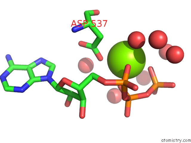
Mono view
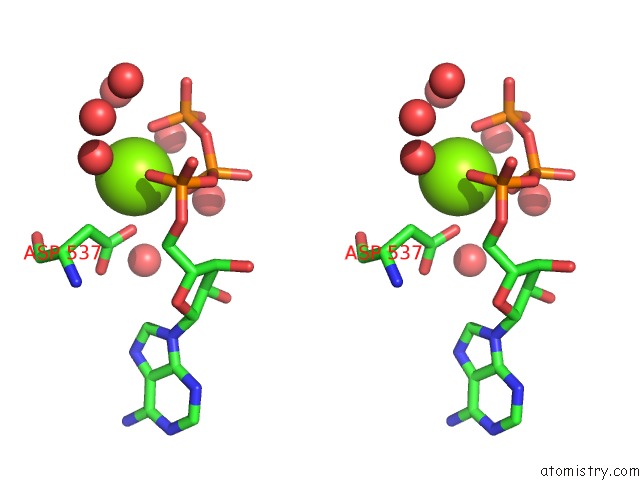
Stereo pair view
Mono view
Stereo pair view
A full contact list of Magnesium with other atoms in the Mg binding
site number 1 of Crystal Structure of Ubiquitin Activating Enzyme 1 (UBA1) in Complex with the Ub E2 UBC4, Ubiquitin, and Atp/Mg within 5.0Å range:
|
Magnesium binding site 2 out of 2 in 4ii2
Go back to
Magnesium binding site 2 out
of 2 in the Crystal Structure of Ubiquitin Activating Enzyme 1 (UBA1) in Complex with the Ub E2 UBC4, Ubiquitin, and Atp/Mg
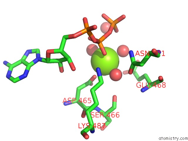
Mono view
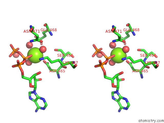
Stereo pair view
Mono view
Stereo pair view
A full contact list of Magnesium with other atoms in the Mg binding
site number 2 of Crystal Structure of Ubiquitin Activating Enzyme 1 (UBA1) in Complex with the Ub E2 UBC4, Ubiquitin, and Atp/Mg within 5.0Å range:
|
Reference:
S.K.Olsen,
C.D.Lima.
Structure of A Ubiquitin E1-E2 Complex: Insights to E1-E2 Thioester Transfer. Mol.Cell V. 49 884 2013.
ISSN: ISSN 1097-2765
PubMed: 23416107
DOI: 10.1016/J.MOLCEL.2013.01.013
Page generated: Fri Aug 16 16:43:27 2024
ISSN: ISSN 1097-2765
PubMed: 23416107
DOI: 10.1016/J.MOLCEL.2013.01.013
Last articles
Mg in 1KZUMg in 1L2X
Mg in 1L3P
Mg in 1L3J
Mg in 1L2O
Mg in 1L2E
Mg in 1L0O
Mg in 1L1R
Mg in 1KXG
Mg in 1KYR