Magnesium »
PDB 4mff-4mpo »
4mif »
Magnesium in PDB 4mif: Pyranose 2-Oxidase From Phanerochaete Chrysosporium, Wild Type From Natural Source
Enzymatic activity of Pyranose 2-Oxidase From Phanerochaete Chrysosporium, Wild Type From Natural Source
All present enzymatic activity of Pyranose 2-Oxidase From Phanerochaete Chrysosporium, Wild Type From Natural Source:
1.1.3.10;
1.1.3.10;
Protein crystallography data
The structure of Pyranose 2-Oxidase From Phanerochaete Chrysosporium, Wild Type From Natural Source, PDB code: 4mif
was solved by
N.Hassan,
T.C.Tan,
O.Spadiut,
I.Pisanelli,
L.Fusco,
D.Haltrich,
C.Peterbauer,
C.Divne,
with X-Ray Crystallography technique. A brief refinement statistics is given in the table below:
| Resolution Low / High (Å) | 46.40 / 1.80 |
| Space group | P 32 2 1 |
| Cell size a, b, c (Å), α, β, γ (°) | 164.034, 164.034, 232.521, 90.00, 90.00, 120.00 |
| R / Rfree (%) | 15.3 / 17.1 |
Magnesium Binding Sites:
The binding sites of Magnesium atom in the Pyranose 2-Oxidase From Phanerochaete Chrysosporium, Wild Type From Natural Source
(pdb code 4mif). This binding sites where shown within
5.0 Angstroms radius around Magnesium atom.
In total only one binding site of Magnesium was determined in the Pyranose 2-Oxidase From Phanerochaete Chrysosporium, Wild Type From Natural Source, PDB code: 4mif:
In total only one binding site of Magnesium was determined in the Pyranose 2-Oxidase From Phanerochaete Chrysosporium, Wild Type From Natural Source, PDB code: 4mif:
Magnesium binding site 1 out of 1 in 4mif
Go back to
Magnesium binding site 1 out
of 1 in the Pyranose 2-Oxidase From Phanerochaete Chrysosporium, Wild Type From Natural Source
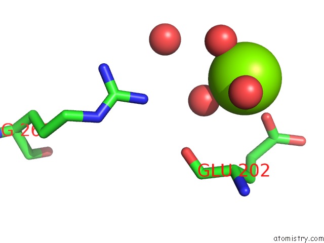
Mono view
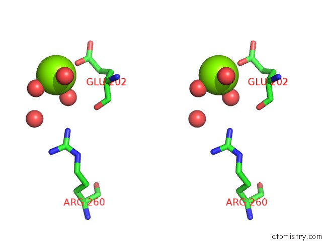
Stereo pair view
Mono view
Stereo pair view
A full contact list of Magnesium with other atoms in the Mg binding
site number 1 of Pyranose 2-Oxidase From Phanerochaete Chrysosporium, Wild Type From Natural Source within 5.0Å range:
|
Reference:
N.Hassan,
T.C.Tan,
O.Spadiut,
I.Pisanelli,
L.Fusco,
D.Haltrich,
C.K.Peterbauer,
C.Divne.
Crystal Structures of Phanerochaete Chrysosporium Pyranose 2-Oxidase Suggest That the N-Terminus Acts As A Propeptide That Assists in Homotetramer Assembly. Febs Open Bio V. 3 496 2013.
ISSN: ESSN 2211-5463
PubMed: 24282677
DOI: 10.1016/J.FOB.2013.10.010
Page generated: Mon Aug 11 20:26:47 2025
ISSN: ESSN 2211-5463
PubMed: 24282677
DOI: 10.1016/J.FOB.2013.10.010
Last articles
Mg in 4Q66Mg in 4Q8B
Mg in 4Q7F
Mg in 4Q5S
Mg in 4Q6X
Mg in 4Q4Z
Mg in 4Q5H
Mg in 4Q4C
Mg in 4Q4D
Mg in 4Q57