Magnesium »
PDB 4osq-4p8r »
4oxd »
Magnesium in PDB 4oxd: Structure of the Ldcb Ld-Carboxypeptidase Reveals the Molecular Basis of Peptidoglycan Recognition
Protein crystallography data
The structure of Structure of the Ldcb Ld-Carboxypeptidase Reveals the Molecular Basis of Peptidoglycan Recognition, PDB code: 4oxd
was solved by
C.N.Hoyland,
C.Aldridge,
R.M.Cleverley,
K.Sidiq,
M.C.Duchene,
R.A.Daniel,
W.Vollmer,
R.J.Lewis,
with X-Ray Crystallography technique. A brief refinement statistics is given in the table below:
| Resolution Low / High (Å) | 47.80 / 2.80 |
| Space group | C 1 2 1 |
| Cell size a, b, c (Å), α, β, γ (°) | 345.954, 42.549, 79.318, 90.00, 93.07, 90.00 |
| R / Rfree (%) | 27.3 / 33.4 |
Other elements in 4oxd:
The structure of Structure of the Ldcb Ld-Carboxypeptidase Reveals the Molecular Basis of Peptidoglycan Recognition also contains other interesting chemical elements:
| Chlorine | (Cl) | 2 atoms |
| Zinc | (Zn) | 18 atoms |
Magnesium Binding Sites:
The binding sites of Magnesium atom in the Structure of the Ldcb Ld-Carboxypeptidase Reveals the Molecular Basis of Peptidoglycan Recognition
(pdb code 4oxd). This binding sites where shown within
5.0 Angstroms radius around Magnesium atom.
In total only one binding site of Magnesium was determined in the Structure of the Ldcb Ld-Carboxypeptidase Reveals the Molecular Basis of Peptidoglycan Recognition, PDB code: 4oxd:
In total only one binding site of Magnesium was determined in the Structure of the Ldcb Ld-Carboxypeptidase Reveals the Molecular Basis of Peptidoglycan Recognition, PDB code: 4oxd:
Magnesium binding site 1 out of 1 in 4oxd
Go back to
Magnesium binding site 1 out
of 1 in the Structure of the Ldcb Ld-Carboxypeptidase Reveals the Molecular Basis of Peptidoglycan Recognition
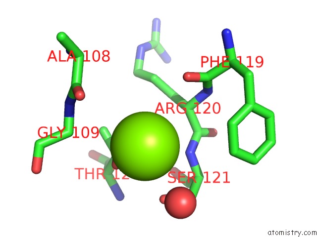
Mono view
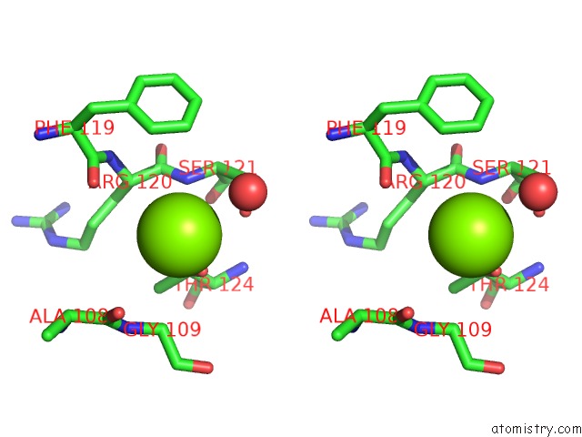
Stereo pair view
Mono view
Stereo pair view
A full contact list of Magnesium with other atoms in the Mg binding
site number 1 of Structure of the Ldcb Ld-Carboxypeptidase Reveals the Molecular Basis of Peptidoglycan Recognition within 5.0Å range:
|
Reference:
C.N.Hoyland,
C.Aldridge,
R.M.Cleverley,
M.C.Duchene,
G.Minasov,
O.Onopriyenko,
K.Sidiq,
P.J.Stogios,
W.F.Anderson,
R.A.Daniel,
A.Savchenko,
W.Vollmer,
R.J.Lewis.
Structure of the Ldcb Ld-Carboxypeptidase Reveals the Molecular Basis of Peptidoglycan Recognition. Structure V. 22 949 2014.
ISSN: ISSN 0969-2126
PubMed: 24909784
DOI: 10.1016/J.STR.2014.04.015
Page generated: Mon Aug 11 21:40:12 2025
ISSN: ISSN 0969-2126
PubMed: 24909784
DOI: 10.1016/J.STR.2014.04.015
Last articles
Mg in 5XNMMg in 5XTM
Mg in 5XUT
Mg in 5XUS
Mg in 5XUJ
Mg in 5XUI
Mg in 5XU1
Mg in 5XT8
Mg in 5XT2
Mg in 5XR7