Magnesium »
PDB 4q8e-4qh0 »
4qan »
Magnesium in PDB 4qan: Crystal Structure of A Hypothetical Protein (RUMGNA_02398) From Ruminococcus Gnavus Atcc 29149 at 2.10 A Resolution
Protein crystallography data
The structure of Crystal Structure of A Hypothetical Protein (RUMGNA_02398) From Ruminococcus Gnavus Atcc 29149 at 2.10 A Resolution, PDB code: 4qan
was solved by
Joint Center For Structural Genomics (Jcsg),
with X-Ray Crystallography technique. A brief refinement statistics is given in the table below:
| Resolution Low / High (Å) | 29.67 / 2.10 |
| Space group | P 21 21 21 |
| Cell size a, b, c (Å), α, β, γ (°) | 51.394, 109.013, 137.307, 90.00, 90.00, 90.00 |
| R / Rfree (%) | 19.1 / 24 |
Magnesium Binding Sites:
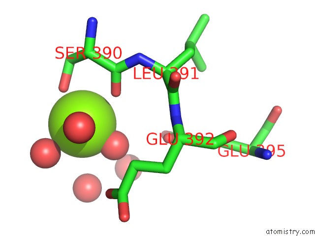
The binding sites of Magnesium atom in the Crystal Structure of A Hypothetical Protein (RUMGNA_02398) From Ruminococcus Gnavus Atcc 29149 at 2.10 A Resolution
(pdb code 4qan). This binding sites where shown within
5.0 Angstroms radius around Magnesium atom.
In total only one binding site of Magnesium was determined in the Crystal Structure of A Hypothetical Protein (RUMGNA_02398) From Ruminococcus Gnavus Atcc 29149 at 2.10 A Resolution, PDB code: 4qan:
In total only one binding site of Magnesium was determined in the Crystal Structure of A Hypothetical Protein (RUMGNA_02398) From Ruminococcus Gnavus Atcc 29149 at 2.10 A Resolution, PDB code: 4qan:
Magnesium binding site 1 out of 1 in 4qan
Go back to
Magnesium binding site 1 out
of 1 in the Crystal Structure of A Hypothetical Protein (RUMGNA_02398) From Ruminococcus Gnavus Atcc 29149 at 2.10 A Resolution
Mono view

Stereo pair view
Mono view
Stereo pair view
A full contact list of Magnesium with other atoms in the Mg binding
site number 1 of Crystal Structure of A Hypothetical Protein (RUMGNA_02398) From Ruminococcus Gnavus Atcc 29149 at 2.10 A Resolution within 5.0Å range:
|
Reference:
Joint Center For Structural Genomics (Jcsg),
Joint Center For Structural Genomics (Jcsg).
N/A N/A.
Page generated: Tue Aug 20 01:52:03 2024
Last articles
Mg in 4JI1Mg in 4JI0
Mg in 4JI2
Mg in 4JI3
Mg in 4JHD
Mg in 4JH6
Mg in 4JH8
Mg in 4JH7
Mg in 4JH3
Mg in 4JH5