Magnesium »
PDB 4rcz-4rkf »
4reu »
Magnesium in PDB 4reu: Revelation of Endogenously Bound FE2+ Ions in the Crystal Structure of Ferritin From Escherichia Coli
Enzymatic activity of Revelation of Endogenously Bound FE2+ Ions in the Crystal Structure of Ferritin From Escherichia Coli
All present enzymatic activity of Revelation of Endogenously Bound FE2+ Ions in the Crystal Structure of Ferritin From Escherichia Coli:
1.16.3.1;
1.16.3.1;
Protein crystallography data
The structure of Revelation of Endogenously Bound FE2+ Ions in the Crystal Structure of Ferritin From Escherichia Coli, PDB code: 4reu
was solved by
V.Thiruselvam,
M.N.Ponnuswamy,
T.S.Kumarevel,
with X-Ray Crystallography technique. A brief refinement statistics is given in the table below:
| Resolution Low / High (Å) | 50.00 / 2.50 |
| Space group | I 4 |
| Cell size a, b, c (Å), α, β, γ (°) | 127.998, 127.998, 170.902, 90.00, 90.00, 90.00 |
| R / Rfree (%) | 17.2 / 23.5 |
Other elements in 4reu:
The structure of Revelation of Endogenously Bound FE2+ Ions in the Crystal Structure of Ferritin From Escherichia Coli also contains other interesting chemical elements:
| Mercury | (Hg) | 2 atoms |
| Iron | (Fe) | 12 atoms |
| Chlorine | (Cl) | 27 atoms |
Magnesium Binding Sites:
The binding sites of Magnesium atom in the Revelation of Endogenously Bound FE2+ Ions in the Crystal Structure of Ferritin From Escherichia Coli
(pdb code 4reu). This binding sites where shown within
5.0 Angstroms radius around Magnesium atom.
In total only one binding site of Magnesium was determined in the Revelation of Endogenously Bound FE2+ Ions in the Crystal Structure of Ferritin From Escherichia Coli, PDB code: 4reu:
In total only one binding site of Magnesium was determined in the Revelation of Endogenously Bound FE2+ Ions in the Crystal Structure of Ferritin From Escherichia Coli, PDB code: 4reu:
Magnesium binding site 1 out of 1 in 4reu
Go back to
Magnesium binding site 1 out
of 1 in the Revelation of Endogenously Bound FE2+ Ions in the Crystal Structure of Ferritin From Escherichia Coli
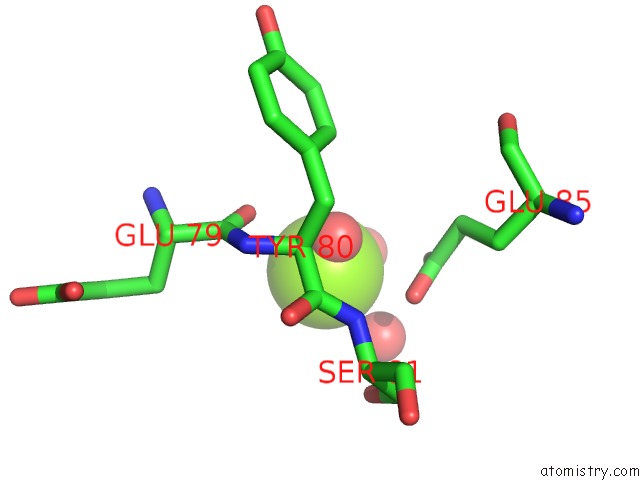
Mono view
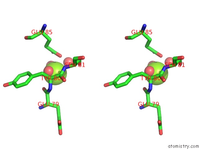
Stereo pair view
Mono view
Stereo pair view
A full contact list of Magnesium with other atoms in the Mg binding
site number 1 of Revelation of Endogenously Bound FE2+ Ions in the Crystal Structure of Ferritin From Escherichia Coli within 5.0Å range:
|
Reference:
V.Thiruselvam,
P.Sivaraman,
T.S.Kumarevel,
M.N.Ponnuswamy.
Revelation of Endogenously Bound Fe(2+) Ions in the Crystal Structure of Ferritin From Escherichia Coli. Biochem.Biophys.Res.Commun. 2014.
ISSN: ESSN 1090-2104
PubMed: 25305494
DOI: 10.1016/J.BBRC.2014.10.007
Page generated: Mon Aug 11 23:16:47 2025
ISSN: ESSN 1090-2104
PubMed: 25305494
DOI: 10.1016/J.BBRC.2014.10.007
Last articles
Mg in 4XSZMg in 4XTJ
Mg in 4XRU
Mg in 4XSY
Mg in 4XSX
Mg in 4XRP
Mg in 4XSH
Mg in 4XSG
Mg in 4XQT
Mg in 4XRH