Magnesium »
PDB 5d2k-5dar »
5dar »
Magnesium in PDB 5dar: Crystal Structure of the Base of the Ribosomal P Stalk From Methanococcus Jannaschii
Protein crystallography data
The structure of Crystal Structure of the Base of the Ribosomal P Stalk From Methanococcus Jannaschii, PDB code: 5dar
was solved by
A.G.Gabdulkhakov,
I.V.Mitroshin,
M.B.Garber,
with X-Ray Crystallography technique. A brief refinement statistics is given in the table below:
| Resolution Low / High (Å) | 20.00 / 2.90 |
| Space group | P 1 21 1 |
| Cell size a, b, c (Å), α, β, γ (°) | 72.396, 88.451, 95.231, 90.00, 102.19, 90.00 |
| R / Rfree (%) | 26.4 / 29.7 |
Other elements in 5dar:
The structure of Crystal Structure of the Base of the Ribosomal P Stalk From Methanococcus Jannaschii also contains other interesting chemical elements:
| Potassium | (K) | 5 atoms |
| Chlorine | (Cl) | 3 atoms |
Magnesium Binding Sites:
The binding sites of Magnesium atom in the Crystal Structure of the Base of the Ribosomal P Stalk From Methanococcus Jannaschii
(pdb code 5dar). This binding sites where shown within
5.0 Angstroms radius around Magnesium atom.
In total 8 binding sites of Magnesium where determined in the Crystal Structure of the Base of the Ribosomal P Stalk From Methanococcus Jannaschii, PDB code: 5dar:
Jump to Magnesium binding site number: 1; 2; 3; 4; 5; 6; 7; 8;
In total 8 binding sites of Magnesium where determined in the Crystal Structure of the Base of the Ribosomal P Stalk From Methanococcus Jannaschii, PDB code: 5dar:
Jump to Magnesium binding site number: 1; 2; 3; 4; 5; 6; 7; 8;
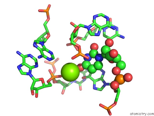
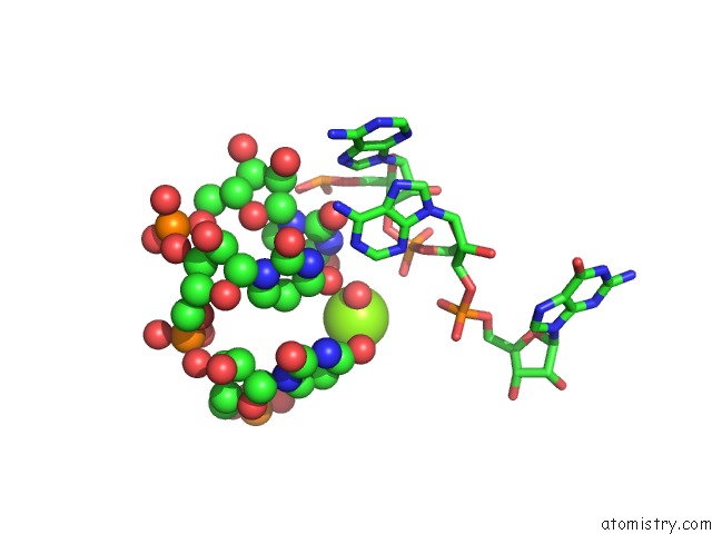
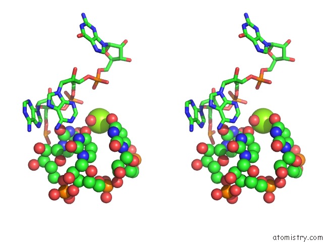
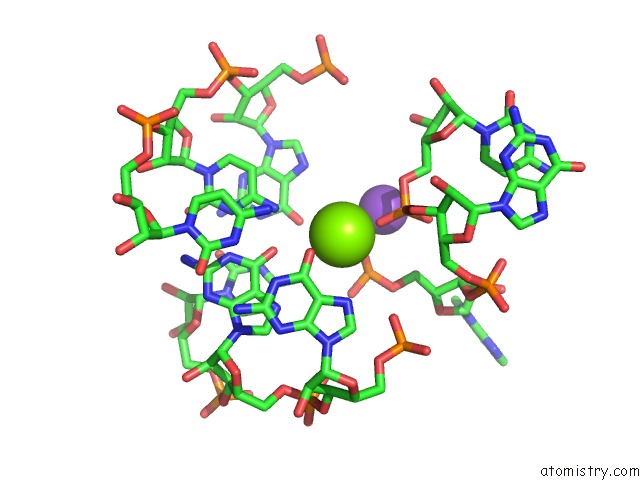
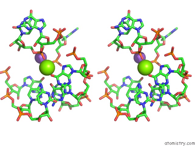
Magnesium binding site 1 out of 8 in 5dar
Go back to
Magnesium binding site 1 out
of 8 in the Crystal Structure of the Base of the Ribosomal P Stalk From Methanococcus Jannaschii
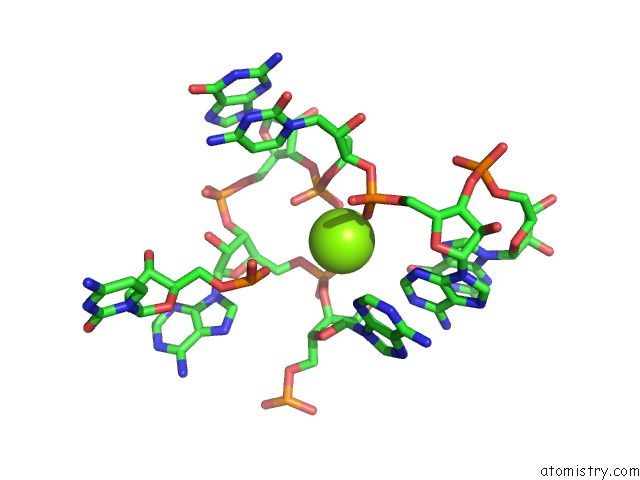
Mono view
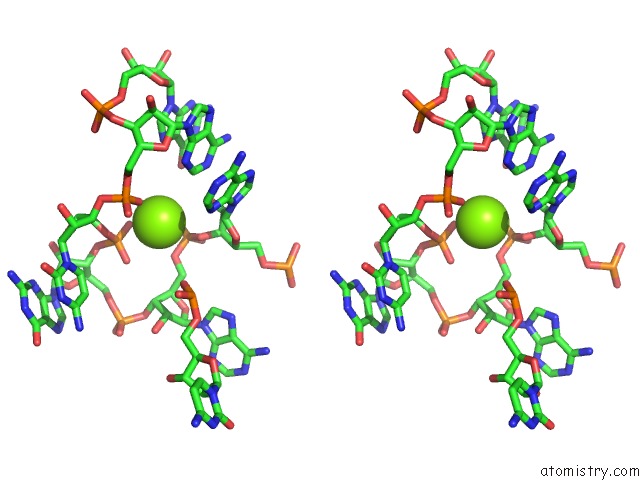
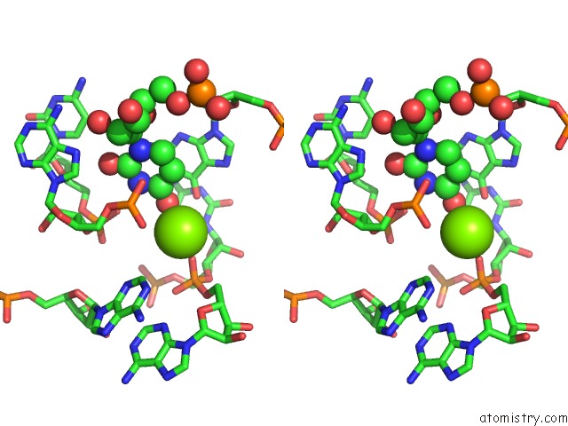
Stereo pair view
Mono view
Stereo pair view
A full contact list of Magnesium with other atoms in the Mg binding
site number 1 of Crystal Structure of the Base of the Ribosomal P Stalk From Methanococcus Jannaschii within 5.0Å range:
|
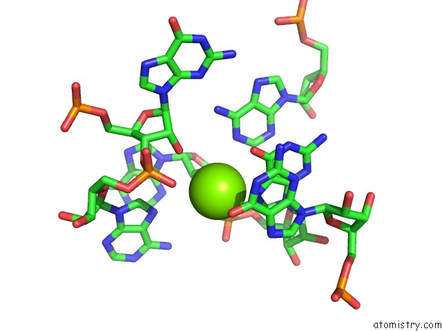
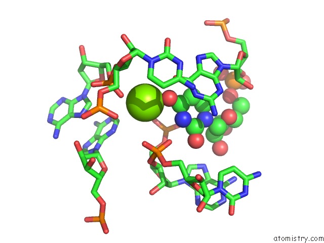
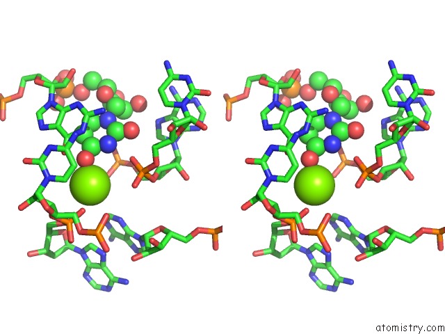
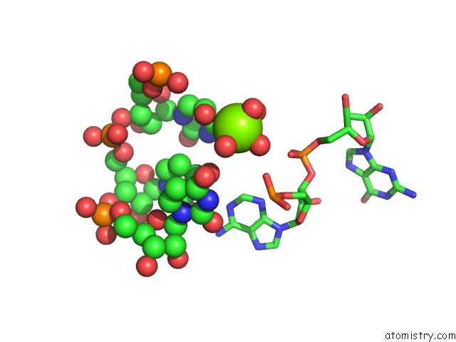
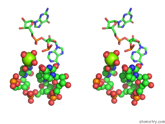
Magnesium binding site 2 out of 8 in 5dar
Go back to
Magnesium binding site 2 out
of 8 in the Crystal Structure of the Base of the Ribosomal P Stalk From Methanococcus Jannaschii
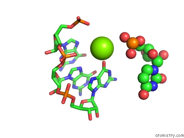
Mono view
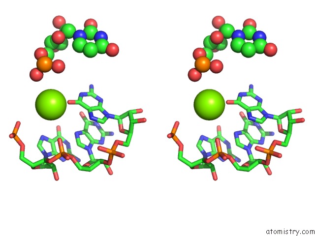
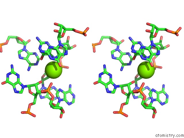
Stereo pair view
Mono view
Stereo pair view
A full contact list of Magnesium with other atoms in the Mg binding
site number 2 of Crystal Structure of the Base of the Ribosomal P Stalk From Methanococcus Jannaschii within 5.0Å range:
|
Magnesium binding site 3 out of 8 in 5dar
Go back to
Magnesium binding site 3 out
of 8 in the Crystal Structure of the Base of the Ribosomal P Stalk From Methanococcus Jannaschii
Mono view
Stereo pair view
Mono view
Stereo pair view
A full contact list of Magnesium with other atoms in the Mg binding
site number 3 of Crystal Structure of the Base of the Ribosomal P Stalk From Methanococcus Jannaschii within 5.0Å range:
|
Magnesium binding site 4 out of 8 in 5dar
Go back to
Magnesium binding site 4 out
of 8 in the Crystal Structure of the Base of the Ribosomal P Stalk From Methanococcus Jannaschii
Mono view
Stereo pair view
Mono view
Stereo pair view
A full contact list of Magnesium with other atoms in the Mg binding
site number 4 of Crystal Structure of the Base of the Ribosomal P Stalk From Methanococcus Jannaschii within 5.0Å range:
|
Magnesium binding site 5 out of 8 in 5dar
Go back to
Magnesium binding site 5 out
of 8 in the Crystal Structure of the Base of the Ribosomal P Stalk From Methanococcus Jannaschii
Mono view
Stereo pair view
Mono view
Stereo pair view
A full contact list of Magnesium with other atoms in the Mg binding
site number 5 of Crystal Structure of the Base of the Ribosomal P Stalk From Methanococcus Jannaschii within 5.0Å range:
|
Magnesium binding site 6 out of 8 in 5dar
Go back to
Magnesium binding site 6 out
of 8 in the Crystal Structure of the Base of the Ribosomal P Stalk From Methanococcus Jannaschii
Mono view
Stereo pair view
Mono view
Stereo pair view
A full contact list of Magnesium with other atoms in the Mg binding
site number 6 of Crystal Structure of the Base of the Ribosomal P Stalk From Methanococcus Jannaschii within 5.0Å range:
|
Magnesium binding site 7 out of 8 in 5dar
Go back to
Magnesium binding site 7 out
of 8 in the Crystal Structure of the Base of the Ribosomal P Stalk From Methanococcus Jannaschii
Mono view
Stereo pair view
Mono view
Stereo pair view
A full contact list of Magnesium with other atoms in the Mg binding
site number 7 of Crystal Structure of the Base of the Ribosomal P Stalk From Methanococcus Jannaschii within 5.0Å range:
|
Magnesium binding site 8 out of 8 in 5dar
Go back to
Magnesium binding site 8 out
of 8 in the Crystal Structure of the Base of the Ribosomal P Stalk From Methanococcus Jannaschii
Mono view
Stereo pair view
Mono view
Stereo pair view
A full contact list of Magnesium with other atoms in the Mg binding
site number 8 of Crystal Structure of the Base of the Ribosomal P Stalk From Methanococcus Jannaschii within 5.0Å range:
|
Reference:
A.G.Gabdulkhakov,
I.V.Mitroshin,
M.B.Garber.
Crystal Structure of the Base of the Ribosomal P Stalk From Methanococcus Jannaschii To Be Published.
Page generated: Tue Aug 12 06:52:03 2025
Last articles
Mg in 6ZXSMg in 7A0Q
Mg in 7A0P
Mg in 7A0C
Mg in 721P
Mg in 6ZXH
Mg in 6ZXG
Mg in 6ZZ6
Mg in 6ZYM
Mg in 6ZY9