Magnesium »
PDB 5ylz-5yw2 »
5ysd »
Magnesium in PDB 5ysd: Crystal Structure of Beta-1,2-Glucooligosaccharide Binding Protein in Complex with Sophorotriose
Protein crystallography data
The structure of Crystal Structure of Beta-1,2-Glucooligosaccharide Binding Protein in Complex with Sophorotriose, PDB code: 5ysd
was solved by
K.Abe,
M.Nakajima,
H.Taguchi,
T.Arakawa,
S.Fushinobu,
with X-Ray Crystallography technique. A brief refinement statistics is given in the table below:
| Resolution Low / High (Å) | 44.95 / 2.10 |
| Space group | P 1 21 1 |
| Cell size a, b, c (Å), α, β, γ (°) | 36.082, 125.561, 91.780, 90.00, 101.61, 90.00 |
| R / Rfree (%) | 16.9 / 20.7 |
Magnesium Binding Sites:
The binding sites of Magnesium atom in the Crystal Structure of Beta-1,2-Glucooligosaccharide Binding Protein in Complex with Sophorotriose
(pdb code 5ysd). This binding sites where shown within
5.0 Angstroms radius around Magnesium atom.
In total only one binding site of Magnesium was determined in the Crystal Structure of Beta-1,2-Glucooligosaccharide Binding Protein in Complex with Sophorotriose, PDB code: 5ysd:
In total only one binding site of Magnesium was determined in the Crystal Structure of Beta-1,2-Glucooligosaccharide Binding Protein in Complex with Sophorotriose, PDB code: 5ysd:
Magnesium binding site 1 out of 1 in 5ysd
Go back to
Magnesium binding site 1 out
of 1 in the Crystal Structure of Beta-1,2-Glucooligosaccharide Binding Protein in Complex with Sophorotriose
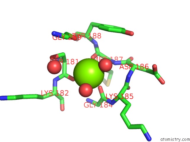
Mono view
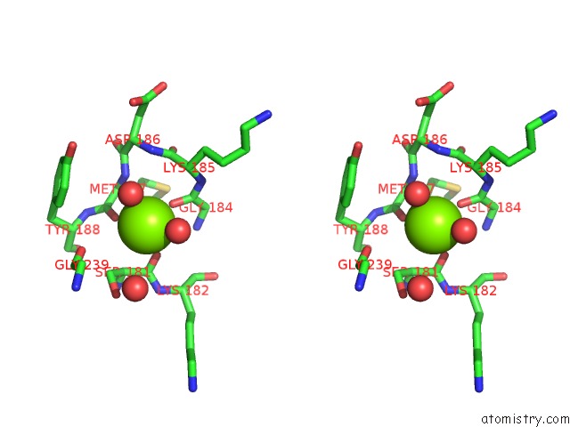
Stereo pair view
Mono view
Stereo pair view
A full contact list of Magnesium with other atoms in the Mg binding
site number 1 of Crystal Structure of Beta-1,2-Glucooligosaccharide Binding Protein in Complex with Sophorotriose within 5.0Å range:
|
Reference:
K.Abe,
N.Sunagawa,
T.Terada,
Y.Takahashi,
T.Arakawa,
K.Igarashi,
M.Samejima,
H.Nakai,
H.Taguchi,
M.Nakajima,
S.Fushinobu.
Structural and Thermodynamic Insights Into Beta-1,2-Glucooligosaccharide Capture By A Solute-Binding Protein Inlisteria Innocua. J. Biol. Chem. V. 293 8812 2018.
ISSN: ESSN 1083-351X
PubMed: 29678880
DOI: 10.1074/JBC.RA117.001536
Page generated: Wed Aug 13 01:03:58 2025
ISSN: ESSN 1083-351X
PubMed: 29678880
DOI: 10.1074/JBC.RA117.001536
Last articles
Mg in 6LU1Mg in 6LT4
Mg in 6LY7
Mg in 6LY6
Mg in 6LY3
Mg in 6LX1
Mg in 6LTW
Mg in 6LVW
Mg in 6LUH
Mg in 6LTS