Magnesium »
PDB 6hns-6hvv »
6ht6 »
Magnesium in PDB 6ht6: Crystal Structure of Glutathione Transferase Omega 2S From Trametes Versicolor in Complex with 2,4-Dihydroxybenzophenone
Protein crystallography data
The structure of Crystal Structure of Glutathione Transferase Omega 2S From Trametes Versicolor in Complex with 2,4-Dihydroxybenzophenone, PDB code: 6ht6
was solved by
M.Schwartz,
F.Favier,
C.Didierjean,
with X-Ray Crystallography technique. A brief refinement statistics is given in the table below:
| Resolution Low / High (Å) | 47.77 / 2.67 |
| Space group | P 21 21 21 |
| Cell size a, b, c (Å), α, β, γ (°) | 58.999, 93.128, 95.548, 90.00, 90.00, 90.00 |
| R / Rfree (%) | 22.1 / 28.4 |
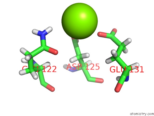
Magnesium Binding Sites:
The binding sites of Magnesium atom in the Crystal Structure of Glutathione Transferase Omega 2S From Trametes Versicolor in Complex with 2,4-Dihydroxybenzophenone
(pdb code 6ht6). This binding sites where shown within
5.0 Angstroms radius around Magnesium atom.
In total only one binding site of Magnesium was determined in the Crystal Structure of Glutathione Transferase Omega 2S From Trametes Versicolor in Complex with 2,4-Dihydroxybenzophenone, PDB code: 6ht6:
In total only one binding site of Magnesium was determined in the Crystal Structure of Glutathione Transferase Omega 2S From Trametes Versicolor in Complex with 2,4-Dihydroxybenzophenone, PDB code: 6ht6:
Magnesium binding site 1 out of 1 in 6ht6
Go back to
Magnesium binding site 1 out
of 1 in the Crystal Structure of Glutathione Transferase Omega 2S From Trametes Versicolor in Complex with 2,4-Dihydroxybenzophenone
Mono view
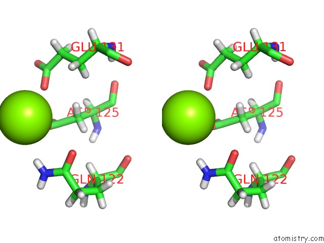
Stereo pair view
Mono view
Stereo pair view
A full contact list of Magnesium with other atoms in the Mg binding
site number 1 of Crystal Structure of Glutathione Transferase Omega 2S From Trametes Versicolor in Complex with 2,4-Dihydroxybenzophenone within 5.0Å range:
|
Reference:
M.Schwartz,
T.Perrot,
E.Aubert,
S.Dumarcay,
F.Favier,
P.Gerardin,
M.Morel-Rouhier,
G.Mulliert,
F.Saiag,
C.Didierjean,
E.Gelhaye.
Molecular Recognition of Wood Polyphenols By Phase II Detoxification Enzymes of the White Rot Trametes Versicolor. Sci Rep V. 8 8472 2018.
ISSN: ESSN 2045-2322
PubMed: 29855494
DOI: 10.1038/S41598-018-26601-3
Page generated: Tue Oct 1 02:13:13 2024
ISSN: ESSN 2045-2322
PubMed: 29855494
DOI: 10.1038/S41598-018-26601-3
Last articles
Mg in 5H1YMg in 5H1B
Mg in 5H1C
Mg in 5H0V
Mg in 5GZA
Mg in 5GZ9
Mg in 5GYN
Mg in 5GXV
Mg in 5GXT
Mg in 5GX5