Magnesium »
PDB 6ugd-6upc »
6uj5 »
Magnesium in PDB 6uj5: Crystal Structure of CAB1 Pantothenate Kinase From Saccharomyces Cerevisiae
Enzymatic activity of Crystal Structure of CAB1 Pantothenate Kinase From Saccharomyces Cerevisiae
All present enzymatic activity of Crystal Structure of CAB1 Pantothenate Kinase From Saccharomyces Cerevisiae:
2.7.1.33;
2.7.1.33;
Protein crystallography data
The structure of Crystal Structure of CAB1 Pantothenate Kinase From Saccharomyces Cerevisiae, PDB code: 6uj5
was solved by
Seattle Structural Genomics Center For Infectious Disease (Ssgcid),
with X-Ray Crystallography technique. A brief refinement statistics is given in the table below:
| Resolution Low / High (Å) | 45.07 / 1.80 |
| Space group | P 1 |
| Cell size a, b, c (Å), α, β, γ (°) | 50.870, 52.750, 71.130, 71.00, 85.59, 64.61 |
| R / Rfree (%) | 16.9 / 20.5 |
Magnesium Binding Sites:
The binding sites of Magnesium atom in the Crystal Structure of CAB1 Pantothenate Kinase From Saccharomyces Cerevisiae
(pdb code 6uj5). This binding sites where shown within
5.0 Angstroms radius around Magnesium atom.
In total only one binding site of Magnesium was determined in the Crystal Structure of CAB1 Pantothenate Kinase From Saccharomyces Cerevisiae, PDB code: 6uj5:
In total only one binding site of Magnesium was determined in the Crystal Structure of CAB1 Pantothenate Kinase From Saccharomyces Cerevisiae, PDB code: 6uj5:
Magnesium binding site 1 out of 1 in 6uj5
Go back to
Magnesium binding site 1 out
of 1 in the Crystal Structure of CAB1 Pantothenate Kinase From Saccharomyces Cerevisiae
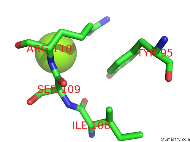
Mono view
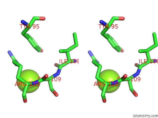
Stereo pair view
Mono view
Stereo pair view
A full contact list of Magnesium with other atoms in the Mg binding
site number 1 of Crystal Structure of CAB1 Pantothenate Kinase From Saccharomyces Cerevisiae within 5.0Å range:
|
Reference:
D.Fox Iii,
P.S.Horanyi,
J.Abendroth,
D.D.Lorimer,
T.E.Edwards.
Crystal Structure of CAB1 Pantothenate Kinase From Saccharomyces Cerevisiae To Be Published.
Page generated: Wed Aug 13 18:34:57 2025
Last articles
Mg in 7JIIMg in 7JIH
Mg in 7JIG
Mg in 7JHO
Mg in 7JIF
Mg in 7JHP
Mg in 7JHL
Mg in 7JH7
Mg in 7JHI
Mg in 7JHN