Magnesium »
PDB 6upd-6uzk »
6uy1 »
Magnesium in PDB 6uy1: Crystal Structure of the STH1 Bromodomain From Saccharomyces Cerevisiae at 2.2 Angstrom Resolution
Enzymatic activity of Crystal Structure of the STH1 Bromodomain From Saccharomyces Cerevisiae at 2.2 Angstrom Resolution
All present enzymatic activity of Crystal Structure of the STH1 Bromodomain From Saccharomyces Cerevisiae at 2.2 Angstrom Resolution:
3.6.4.12;
3.6.4.12;
Protein crystallography data
The structure of Crystal Structure of the STH1 Bromodomain From Saccharomyces Cerevisiae at 2.2 Angstrom Resolution, PDB code: 6uy1
was solved by
P.Stavropoulos,
A.Hoelz,
with X-Ray Crystallography technique. A brief refinement statistics is given in the table below:
| Resolution Low / High (Å) | 19.95 / 2.21 |
| Space group | P 1 21 1 |
| Cell size a, b, c (Å), α, β, γ (°) | 76.279, 76.281, 98.443, 90.00, 96.65, 90.00 |
| R / Rfree (%) | 18.4 / 22.8 |
Magnesium Binding Sites:
The binding sites of Magnesium atom in the Crystal Structure of the STH1 Bromodomain From Saccharomyces Cerevisiae at 2.2 Angstrom Resolution
(pdb code 6uy1). This binding sites where shown within
5.0 Angstroms radius around Magnesium atom.
In total only one binding site of Magnesium was determined in the Crystal Structure of the STH1 Bromodomain From Saccharomyces Cerevisiae at 2.2 Angstrom Resolution, PDB code: 6uy1:
In total only one binding site of Magnesium was determined in the Crystal Structure of the STH1 Bromodomain From Saccharomyces Cerevisiae at 2.2 Angstrom Resolution, PDB code: 6uy1:
Magnesium binding site 1 out of 1 in 6uy1
Go back to
Magnesium binding site 1 out
of 1 in the Crystal Structure of the STH1 Bromodomain From Saccharomyces Cerevisiae at 2.2 Angstrom Resolution
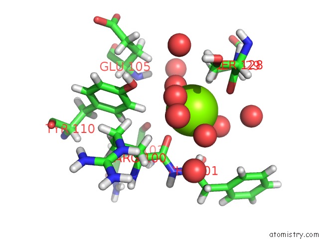
Mono view
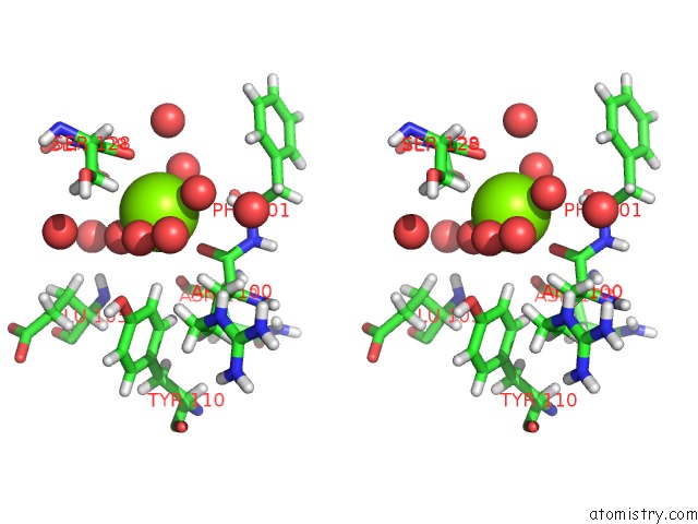
Stereo pair view
Mono view
Stereo pair view
A full contact list of Magnesium with other atoms in the Mg binding
site number 1 of Crystal Structure of the STH1 Bromodomain From Saccharomyces Cerevisiae at 2.2 Angstrom Resolution within 5.0Å range:
|
Reference:
P.Stavropoulos,
A.Hoelz.
Crystal Structure of the STH1 Bromodomain From Saccharomyces Cerevisiae at 2.2 Angstrom Resolution To Be Published.
Page generated: Wed Aug 13 18:45:43 2025
Last articles
Mg in 7M4UMg in 7M5Y
Mg in 7M6J
Mg in 7M5X
Mg in 7M5V
Mg in 7M4E
Mg in 7M4F
Mg in 7M4G
Mg in 7M45
Mg in 7M46