Magnesium »
PDB 7k16-7kee »
7k72 »
Magnesium in PDB 7k72: Structure of Dna Ligase A From Mycobacterium Tuberculosis Bound to Nad
Enzymatic activity of Structure of Dna Ligase A From Mycobacterium Tuberculosis Bound to Nad
All present enzymatic activity of Structure of Dna Ligase A From Mycobacterium Tuberculosis Bound to Nad:
6.5.1.2;
6.5.1.2;
Protein crystallography data
The structure of Structure of Dna Ligase A From Mycobacterium Tuberculosis Bound to Nad, PDB code: 7k72
was solved by
Seattle Structural Genomics Center For Infectious Disease (Ssgcid),
with X-Ray Crystallography technique. A brief refinement statistics is given in the table below:
| Resolution Low / High (Å) | 44.59 / 2.05 |
| Space group | P 1 |
| Cell size a, b, c (Å), α, β, γ (°) | 50.940, 63.810, 120.730, 96.22, 89.55, 111.97 |
| R / Rfree (%) | 16.8 / 20.6 |
Other elements in 7k72:
The structure of Structure of Dna Ligase A From Mycobacterium Tuberculosis Bound to Nad also contains other interesting chemical elements:
| Calcium | (Ca) | 2 atoms |
Magnesium Binding Sites:
The binding sites of Magnesium atom in the Structure of Dna Ligase A From Mycobacterium Tuberculosis Bound to Nad
(pdb code 7k72). This binding sites where shown within
5.0 Angstroms radius around Magnesium atom.
In total 9 binding sites of Magnesium where determined in the Structure of Dna Ligase A From Mycobacterium Tuberculosis Bound to Nad, PDB code: 7k72:
Jump to Magnesium binding site number: 1; 2; 3; 4; 5; 6; 7; 8; 9;
In total 9 binding sites of Magnesium where determined in the Structure of Dna Ligase A From Mycobacterium Tuberculosis Bound to Nad, PDB code: 7k72:
Jump to Magnesium binding site number: 1; 2; 3; 4; 5; 6; 7; 8; 9;
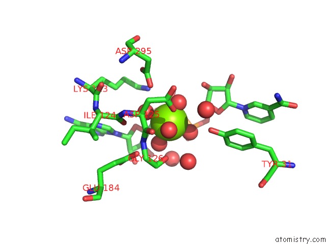
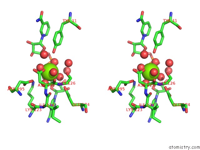
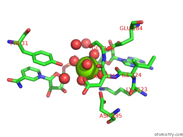
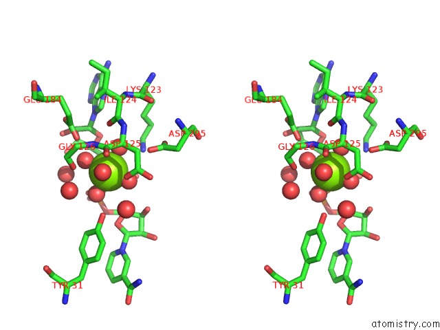
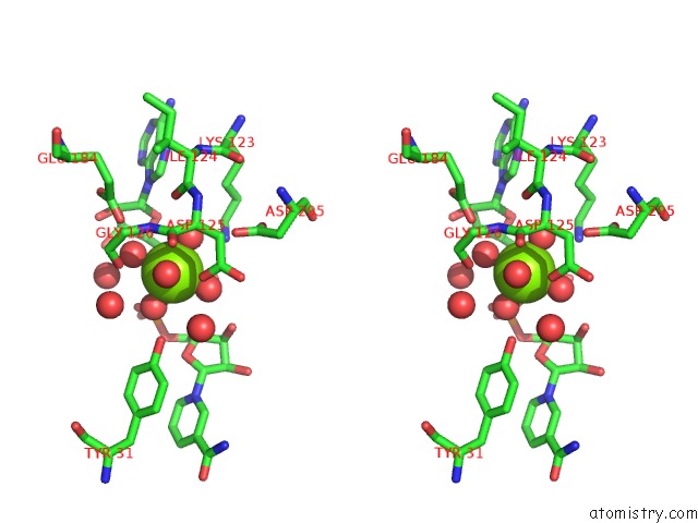
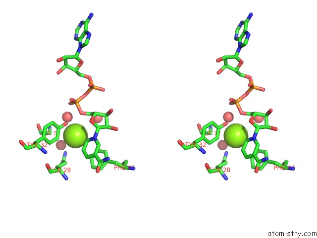
Magnesium binding site 1 out of 9 in 7k72
Go back to
Magnesium binding site 1 out
of 9 in the Structure of Dna Ligase A From Mycobacterium Tuberculosis Bound to Nad
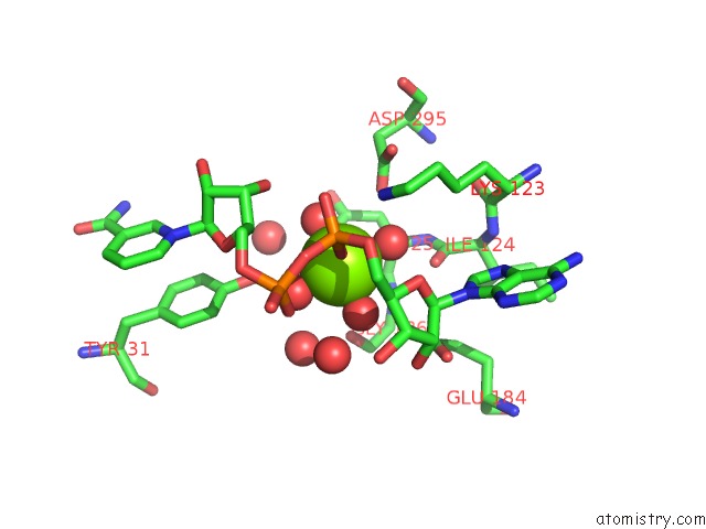
Mono view
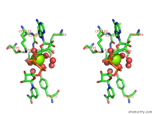
Stereo pair view
Mono view
Stereo pair view
A full contact list of Magnesium with other atoms in the Mg binding
site number 1 of Structure of Dna Ligase A From Mycobacterium Tuberculosis Bound to Nad within 5.0Å range:
|
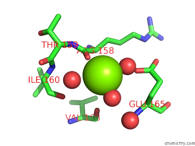
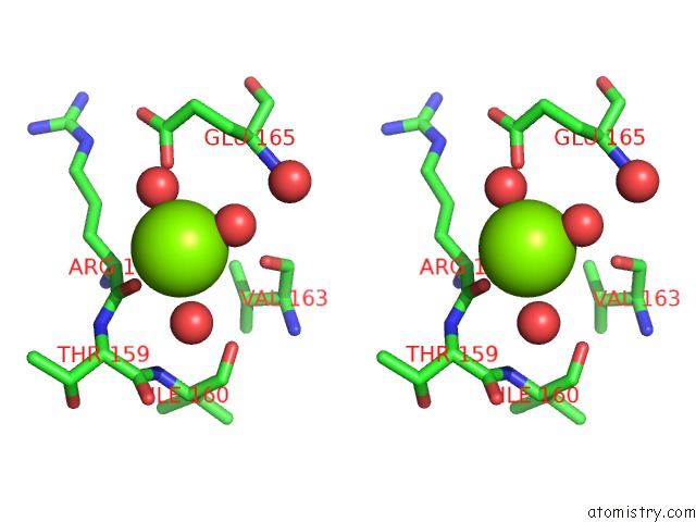
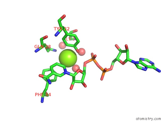
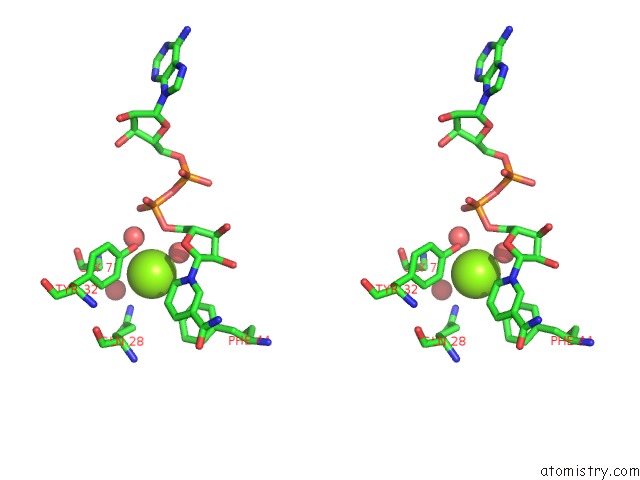
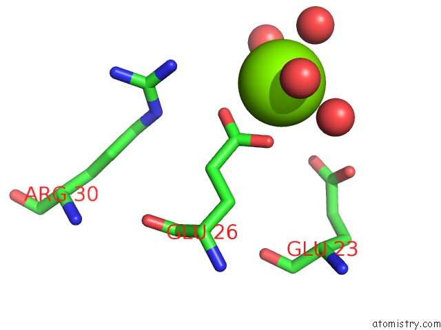
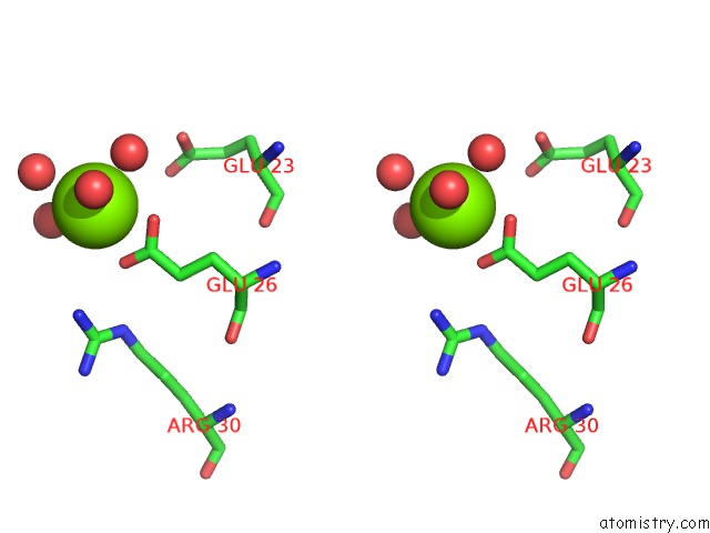
Magnesium binding site 2 out of 9 in 7k72
Go back to
Magnesium binding site 2 out
of 9 in the Structure of Dna Ligase A From Mycobacterium Tuberculosis Bound to Nad
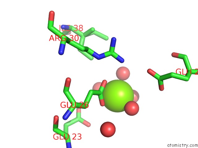
Mono view
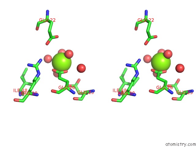
Stereo pair view
Mono view
Stereo pair view
A full contact list of Magnesium with other atoms in the Mg binding
site number 2 of Structure of Dna Ligase A From Mycobacterium Tuberculosis Bound to Nad within 5.0Å range:
|
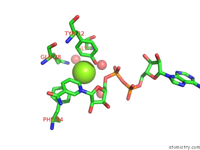
Magnesium binding site 3 out of 9 in 7k72
Go back to
Magnesium binding site 3 out
of 9 in the Structure of Dna Ligase A From Mycobacterium Tuberculosis Bound to Nad
Mono view
Stereo pair view
Mono view
Stereo pair view
A full contact list of Magnesium with other atoms in the Mg binding
site number 3 of Structure of Dna Ligase A From Mycobacterium Tuberculosis Bound to Nad within 5.0Å range:
|
Magnesium binding site 4 out of 9 in 7k72
Go back to
Magnesium binding site 4 out
of 9 in the Structure of Dna Ligase A From Mycobacterium Tuberculosis Bound to Nad
Mono view
Stereo pair view
Mono view
Stereo pair view
A full contact list of Magnesium with other atoms in the Mg binding
site number 4 of Structure of Dna Ligase A From Mycobacterium Tuberculosis Bound to Nad within 5.0Å range:
|
Magnesium binding site 5 out of 9 in 7k72
Go back to
Magnesium binding site 5 out
of 9 in the Structure of Dna Ligase A From Mycobacterium Tuberculosis Bound to Nad
Mono view
Stereo pair view
Mono view
Stereo pair view
A full contact list of Magnesium with other atoms in the Mg binding
site number 5 of Structure of Dna Ligase A From Mycobacterium Tuberculosis Bound to Nad within 5.0Å range:
|
Magnesium binding site 6 out of 9 in 7k72
Go back to
Magnesium binding site 6 out
of 9 in the Structure of Dna Ligase A From Mycobacterium Tuberculosis Bound to Nad
Mono view
Stereo pair view
Mono view
Stereo pair view
A full contact list of Magnesium with other atoms in the Mg binding
site number 6 of Structure of Dna Ligase A From Mycobacterium Tuberculosis Bound to Nad within 5.0Å range:
|
Magnesium binding site 7 out of 9 in 7k72
Go back to
Magnesium binding site 7 out
of 9 in the Structure of Dna Ligase A From Mycobacterium Tuberculosis Bound to Nad

Mono view
Stereo pair view
Mono view
Stereo pair view
A full contact list of Magnesium with other atoms in the Mg binding
site number 7 of Structure of Dna Ligase A From Mycobacterium Tuberculosis Bound to Nad within 5.0Å range:
|
Magnesium binding site 8 out of 9 in 7k72
Go back to
Magnesium binding site 8 out
of 9 in the Structure of Dna Ligase A From Mycobacterium Tuberculosis Bound to Nad
Mono view
Stereo pair view
Mono view
Stereo pair view
A full contact list of Magnesium with other atoms in the Mg binding
site number 8 of Structure of Dna Ligase A From Mycobacterium Tuberculosis Bound to Nad within 5.0Å range:
|
Magnesium binding site 9 out of 9 in 7k72
Go back to
Magnesium binding site 9 out
of 9 in the Structure of Dna Ligase A From Mycobacterium Tuberculosis Bound to Nad
Mono view
Stereo pair view
Mono view
Stereo pair view
A full contact list of Magnesium with other atoms in the Mg binding
site number 9 of Structure of Dna Ligase A From Mycobacterium Tuberculosis Bound to Nad within 5.0Å range:
|
Reference:
S.L.Delker,
J.Abendroth,
D.D.Lorimer,
P.S.Horanyi,
T.E.Edwards.
Structure of Dna Ligase A From Mycobacterium Tuberculosis Bound to Nad To Be Published.
Page generated: Wed Oct 2 22:09:23 2024
Last articles
Mg in 4Q39Mg in 4Q2G
Mg in 4Q2E
Mg in 4Q23
Mg in 4Q15
Mg in 4Q1V
Mg in 4Q2D
Mg in 4Q21
Mg in 4Q01
Mg in 4Q04