Magnesium »
PDB 8f8f-8fi2 »
8fdb »
Magnesium in PDB 8fdb: Crystal Structure of Nagb-II Phosphosugar Isomerase From Shewanella Denitrificans OS217 in Complex with Glucitolamine-6-Phosphate at 3.06 A Resolution.
Protein crystallography data
The structure of Crystal Structure of Nagb-II Phosphosugar Isomerase From Shewanella Denitrificans OS217 in Complex with Glucitolamine-6-Phosphate at 3.06 A Resolution., PDB code: 8fdb
was solved by
A.Rodriguez-Romero,
A.Rodriguez-Hernandez,
J.Marcos-Viquez,
I.Bustos-Jaimes,
with X-Ray Crystallography technique. A brief refinement statistics is given in the table below:
| Resolution Low / High (Å) | 34.95 / 3.06 |
| Space group | C 2 2 21 |
| Cell size a, b, c (Å), α, β, γ (°) | 71.885, 113.142, 171.008, 90, 90, 90 |
| R / Rfree (%) | 20.6 / 24.2 |
Magnesium Binding Sites:
The binding sites of Magnesium atom in the Crystal Structure of Nagb-II Phosphosugar Isomerase From Shewanella Denitrificans OS217 in Complex with Glucitolamine-6-Phosphate at 3.06 A Resolution.
(pdb code 8fdb). This binding sites where shown within
5.0 Angstroms radius around Magnesium atom.
In total 2 binding sites of Magnesium where determined in the Crystal Structure of Nagb-II Phosphosugar Isomerase From Shewanella Denitrificans OS217 in Complex with Glucitolamine-6-Phosphate at 3.06 A Resolution., PDB code: 8fdb:
Jump to Magnesium binding site number: 1; 2;
In total 2 binding sites of Magnesium where determined in the Crystal Structure of Nagb-II Phosphosugar Isomerase From Shewanella Denitrificans OS217 in Complex with Glucitolamine-6-Phosphate at 3.06 A Resolution., PDB code: 8fdb:
Jump to Magnesium binding site number: 1; 2;
Magnesium binding site 1 out of 2 in 8fdb
Go back to
Magnesium binding site 1 out
of 2 in the Crystal Structure of Nagb-II Phosphosugar Isomerase From Shewanella Denitrificans OS217 in Complex with Glucitolamine-6-Phosphate at 3.06 A Resolution.

Mono view
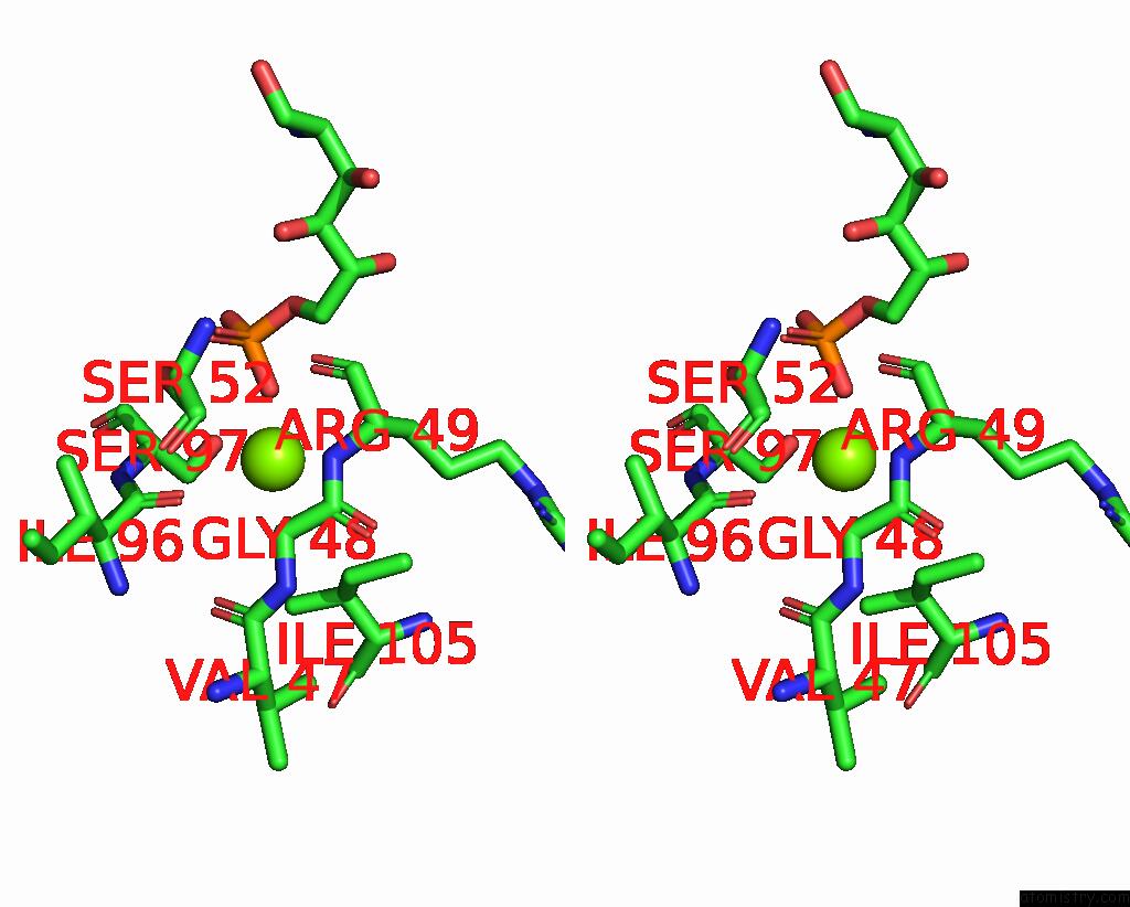
Stereo pair view
Mono view
Stereo pair view
A full contact list of Magnesium with other atoms in the Mg binding
site number 1 of Crystal Structure of Nagb-II Phosphosugar Isomerase From Shewanella Denitrificans OS217 in Complex with Glucitolamine-6-Phosphate at 3.06 A Resolution. within 5.0Å range:
|
Magnesium binding site 2 out of 2 in 8fdb
Go back to
Magnesium binding site 2 out
of 2 in the Crystal Structure of Nagb-II Phosphosugar Isomerase From Shewanella Denitrificans OS217 in Complex with Glucitolamine-6-Phosphate at 3.06 A Resolution.

Mono view
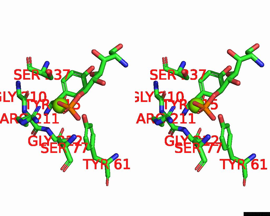
Stereo pair view
Mono view
Stereo pair view
A full contact list of Magnesium with other atoms in the Mg binding
site number 2 of Crystal Structure of Nagb-II Phosphosugar Isomerase From Shewanella Denitrificans OS217 in Complex with Glucitolamine-6-Phosphate at 3.06 A Resolution. within 5.0Å range:
|
Reference:
J.Marcos-Viquez,
A.Rodriguez-Hernandez,
L.I.Alvarez-Anorve,
A.Medina-Garcia,
J.Plumbridge,
M.L.Calcagno,
A.Rodriguez-Romero,
I.Bustos-Jaimes.
Substrate Binding in the Allosteric Site Mimics Homotropic Cooperativity in the Sis-Fold Glucosamine-6-Phosphate Deaminases. Protein Sci. E4651 2023.
ISSN: ESSN 1469-896X
PubMed: 37145875
DOI: 10.1002/PRO.4651
Page generated: Fri Oct 4 02:29:13 2024
ISSN: ESSN 1469-896X
PubMed: 37145875
DOI: 10.1002/PRO.4651
Last articles
Mg in 7DUJMg in 7DUI
Mg in 7DUH
Mg in 7DUG
Mg in 7DR0
Mg in 7DR1
Mg in 7DU2
Mg in 7DSP
Mg in 7DSJ
Mg in 7DSI