Magnesium »
PDB 8p0g-8p5u »
8p5s »
Magnesium in PDB 8p5s: Crystal Structure of the Homohexameric 2-Oxoglutarate Dehydrogenase Odha From Corynebacterium Glutamicum
Enzymatic activity of Crystal Structure of the Homohexameric 2-Oxoglutarate Dehydrogenase Odha From Corynebacterium Glutamicum
All present enzymatic activity of Crystal Structure of the Homohexameric 2-Oxoglutarate Dehydrogenase Odha From Corynebacterium Glutamicum:
1.2.4.2; 2.3.1.61;
1.2.4.2; 2.3.1.61;
Protein crystallography data
The structure of Crystal Structure of the Homohexameric 2-Oxoglutarate Dehydrogenase Odha From Corynebacterium Glutamicum, PDB code: 8p5s
was solved by
L.Yang,
A.Boyko,
M.Bellinzoni,
with X-Ray Crystallography technique. A brief refinement statistics is given in the table below:
| Resolution Low / High (Å) | 27.53 / 2.46 |
| Space group | H 3 2 |
| Cell size a, b, c (Å), α, β, γ (°) | 150.987, 150.987, 314.335, 90, 90, 120 |
| R / Rfree (%) | 20.4 / 25.1 |
Magnesium Binding Sites:
The binding sites of Magnesium atom in the Crystal Structure of the Homohexameric 2-Oxoglutarate Dehydrogenase Odha From Corynebacterium Glutamicum
(pdb code 8p5s). This binding sites where shown within
5.0 Angstroms radius around Magnesium atom.
In total only one binding site of Magnesium was determined in the Crystal Structure of the Homohexameric 2-Oxoglutarate Dehydrogenase Odha From Corynebacterium Glutamicum, PDB code: 8p5s:
In total only one binding site of Magnesium was determined in the Crystal Structure of the Homohexameric 2-Oxoglutarate Dehydrogenase Odha From Corynebacterium Glutamicum, PDB code: 8p5s:
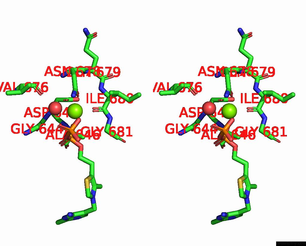
Magnesium binding site 1 out of 1 in 8p5s
Go back to
Magnesium binding site 1 out
of 1 in the Crystal Structure of the Homohexameric 2-Oxoglutarate Dehydrogenase Odha From Corynebacterium Glutamicum

Mono view
Stereo pair view
Mono view
Stereo pair view
A full contact list of Magnesium with other atoms in the Mg binding
site number 1 of Crystal Structure of the Homohexameric 2-Oxoglutarate Dehydrogenase Odha From Corynebacterium Glutamicum within 5.0Å range:
|
Reference:
L.Yang,
T.Wagner,
A.Mechaly,
A.Boyko,
E.M.Bruch,
D.Megrian,
F.Gubellini,
P.M.Alzari,
M.Bellinzoni.
High Resolution Cryo-Em and Crystallographic Snapshots of the Actinobacterial Two-in-One 2-Oxoglutarate Dehydrogenase Nat Commun 2023.
ISSN: ESSN 2041-1723
DOI: 10.1038/S41467-023-40253-6
Page generated: Fri Oct 4 15:52:25 2024
ISSN: ESSN 2041-1723
DOI: 10.1038/S41467-023-40253-6
Last articles
Mg in 4Q39Mg in 4Q2G
Mg in 4Q2E
Mg in 4Q23
Mg in 4Q15
Mg in 4Q1V
Mg in 4Q2D
Mg in 4Q21
Mg in 4Q01
Mg in 4Q04