Magnesium »
PDB 2a6h-2alz »
2ajp »
Magnesium in PDB 2ajp: Crystal Structure of A Human Pyridoxal Kinase
Enzymatic activity of Crystal Structure of A Human Pyridoxal Kinase
All present enzymatic activity of Crystal Structure of A Human Pyridoxal Kinase:
2.7.1.35;
2.7.1.35;
Protein crystallography data
The structure of Crystal Structure of A Human Pyridoxal Kinase, PDB code: 2ajp
was solved by
S.Ismail,
S.Dimov,
A.Atanassova,
W.Tempel,
C.Arrowsmith,
A.Edwards,
M.Sundstrom,
J.Weigelt,
A.Bochkarev,
H.Park,
Structural Genomicsconsortium (Sgc),
with X-Ray Crystallography technique. A brief refinement statistics is given in the table below:
| Resolution Low / High (Å) | 46.08 / 2.50 |
| Space group | I 2 2 2 |
| Cell size a, b, c (Å), α, β, γ (°) | 92.132, 114.971, 169.261, 90.00, 90.00, 90.00 |
| R / Rfree (%) | 24.1 / 28.3 |
Magnesium Binding Sites:
The binding sites of Magnesium atom in the Crystal Structure of A Human Pyridoxal Kinase
(pdb code 2ajp). This binding sites where shown within
5.0 Angstroms radius around Magnesium atom.
In total 2 binding sites of Magnesium where determined in the Crystal Structure of A Human Pyridoxal Kinase, PDB code: 2ajp:
Jump to Magnesium binding site number: 1; 2;
In total 2 binding sites of Magnesium where determined in the Crystal Structure of A Human Pyridoxal Kinase, PDB code: 2ajp:
Jump to Magnesium binding site number: 1; 2;
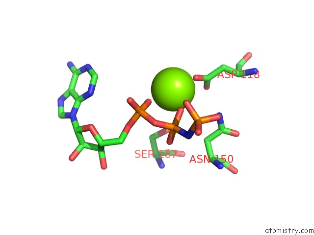
Magnesium binding site 1 out of 2 in 2ajp
Go back to
Magnesium binding site 1 out
of 2 in the Crystal Structure of A Human Pyridoxal Kinase
Mono view

Stereo pair view
Mono view
Stereo pair view
A full contact list of Magnesium with other atoms in the Mg binding
site number 1 of Crystal Structure of A Human Pyridoxal Kinase within 5.0Å range:
|
Magnesium binding site 2 out of 2 in 2ajp
Go back to
Magnesium binding site 2 out
of 2 in the Crystal Structure of A Human Pyridoxal Kinase

Mono view

Stereo pair view
Mono view
Stereo pair view
A full contact list of Magnesium with other atoms in the Mg binding
site number 2 of Crystal Structure of A Human Pyridoxal Kinase within 5.0Å range:
|
Reference:
S.Ismail,
S.Dimov,
A.Atanassova,
W.Tempel,
C.Arrowsmith,
A.Edwards,
M.Sundstrom,
J.Weigelt,
A.Bochkarev,
H.Park,
Structural Genomics Consortium (Sgc).
Crystal Structure of A Human Pyridoxal Kinase To Be Published.
Page generated: Sun Aug 10 09:47:10 2025
Last articles
Mg in 6ZUPMg in 6ZTC
Mg in 6ZT3
Mg in 6ZSI
Mg in 6ZSJ
Mg in 6ZR5
Mg in 6ZRN
Mg in 6ZQZ
Mg in 6ZQN
Mg in 6ZQT