Magnesium »
PDB 2bt6-2c3y »
2c3y »
Magnesium in PDB 2c3y: Crystal Structure of the Radical Form of Pyruvate:Ferredoxin Oxidoreductase From Desulfovibrio Africanus
Enzymatic activity of Crystal Structure of the Radical Form of Pyruvate:Ferredoxin Oxidoreductase From Desulfovibrio Africanus
All present enzymatic activity of Crystal Structure of the Radical Form of Pyruvate:Ferredoxin Oxidoreductase From Desulfovibrio Africanus:
1.2.7.1;
1.2.7.1;
Protein crystallography data
The structure of Crystal Structure of the Radical Form of Pyruvate:Ferredoxin Oxidoreductase From Desulfovibrio Africanus, PDB code: 2c3y
was solved by
C.Cavazza,
C.Contreras-Martel,
L.Pieulle,
E.Chabriere,
E.C.Hatchikian,
J.C.Fontecilla-Camps,
with X-Ray Crystallography technique. A brief refinement statistics is given in the table below:
| Resolution Low / High (Å) | 8.00 / 1.93 |
| Space group | P 21 21 21 |
| Cell size a, b, c (Å), α, β, γ (°) | 86.081, 145.639, 211.619, 90.00, 90.00, 90.00 |
| R / Rfree (%) | 18.6 / 22.8 |
Other elements in 2c3y:
The structure of Crystal Structure of the Radical Form of Pyruvate:Ferredoxin Oxidoreductase From Desulfovibrio Africanus also contains other interesting chemical elements:
| Iron | (Fe) | 24 atoms |
| Calcium | (Ca) | 2 atoms |
Magnesium Binding Sites:
The binding sites of Magnesium atom in the Crystal Structure of the Radical Form of Pyruvate:Ferredoxin Oxidoreductase From Desulfovibrio Africanus
(pdb code 2c3y). This binding sites where shown within
5.0 Angstroms radius around Magnesium atom.
In total 2 binding sites of Magnesium where determined in the Crystal Structure of the Radical Form of Pyruvate:Ferredoxin Oxidoreductase From Desulfovibrio Africanus, PDB code: 2c3y:
Jump to Magnesium binding site number: 1; 2;
In total 2 binding sites of Magnesium where determined in the Crystal Structure of the Radical Form of Pyruvate:Ferredoxin Oxidoreductase From Desulfovibrio Africanus, PDB code: 2c3y:
Jump to Magnesium binding site number: 1; 2;
Magnesium binding site 1 out of 2 in 2c3y
Go back to
Magnesium binding site 1 out
of 2 in the Crystal Structure of the Radical Form of Pyruvate:Ferredoxin Oxidoreductase From Desulfovibrio Africanus
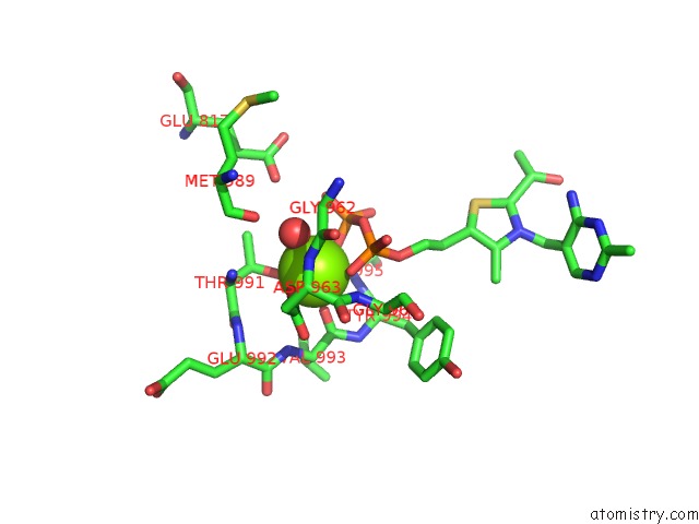
Mono view
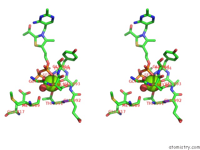
Stereo pair view
Mono view
Stereo pair view
A full contact list of Magnesium with other atoms in the Mg binding
site number 1 of Crystal Structure of the Radical Form of Pyruvate:Ferredoxin Oxidoreductase From Desulfovibrio Africanus within 5.0Å range:
|
Magnesium binding site 2 out of 2 in 2c3y
Go back to
Magnesium binding site 2 out
of 2 in the Crystal Structure of the Radical Form of Pyruvate:Ferredoxin Oxidoreductase From Desulfovibrio Africanus
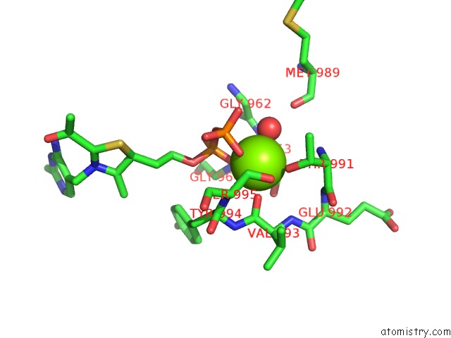
Mono view
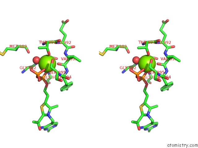
Stereo pair view
Mono view
Stereo pair view
A full contact list of Magnesium with other atoms in the Mg binding
site number 2 of Crystal Structure of the Radical Form of Pyruvate:Ferredoxin Oxidoreductase From Desulfovibrio Africanus within 5.0Å range:
|
Reference:
C.Cavazza,
C.Contreras-Martel,
L.Pieulle,
E.Chabriere,
E.C.Hatchikian,
J.C.Fontecilla-Camps.
Flexibility of Thiamine Diphosphate Revealed By Kinetic Crystallographic Studies of the Reaction of Pyruvate-Ferredoxin Oxidoreductase with Pyruvate. Structure V. 14 217 2006.
ISSN: ISSN 0969-2126
PubMed: 16472741
DOI: 10.1016/J.STR.2005.10.013
Page generated: Sun Aug 10 10:13:38 2025
ISSN: ISSN 0969-2126
PubMed: 16472741
DOI: 10.1016/J.STR.2005.10.013
Last articles
Mg in 5ZKJMg in 5ZKI
Mg in 5ZK6
Mg in 5ZE9
Mg in 5ZFX
Mg in 5ZCT
Mg in 5ZE6
Mg in 5ZE4
Mg in 5ZDN
Mg in 5ZE0