Magnesium »
PDB 2c42-2cic »
2cfs »
Magnesium in PDB 2cfs: Crystal Structure of Human Pyridoxal 5'-Phosphate Phosphatase
Enzymatic activity of Crystal Structure of Human Pyridoxal 5'-Phosphate Phosphatase
All present enzymatic activity of Crystal Structure of Human Pyridoxal 5'-Phosphate Phosphatase:
3.1.3.74;
3.1.3.74;
Protein crystallography data
The structure of Crystal Structure of Human Pyridoxal 5'-Phosphate Phosphatase, PDB code: 2cfs
was solved by
B.S.Kang,
H.J.Cho,
K.J.Kim,
O.S.Kwon,
with X-Ray Crystallography technique. A brief refinement statistics is given in the table below:
| Resolution Low / High (Å) | 18.64 / 2.4 |
| Space group | P 43 21 2 |
| Cell size a, b, c (Å), α, β, γ (°) | 54.631, 54.631, 213.985, 90.00, 90.00, 90.00 |
| R / Rfree (%) | 20.7 / 25.7 |
Magnesium Binding Sites:
The binding sites of Magnesium atom in the Crystal Structure of Human Pyridoxal 5'-Phosphate Phosphatase
(pdb code 2cfs). This binding sites where shown within
5.0 Angstroms radius around Magnesium atom.
In total 2 binding sites of Magnesium where determined in the Crystal Structure of Human Pyridoxal 5'-Phosphate Phosphatase, PDB code: 2cfs:
Jump to Magnesium binding site number: 1; 2;
In total 2 binding sites of Magnesium where determined in the Crystal Structure of Human Pyridoxal 5'-Phosphate Phosphatase, PDB code: 2cfs:
Jump to Magnesium binding site number: 1; 2;
Magnesium binding site 1 out of 2 in 2cfs
Go back to
Magnesium binding site 1 out
of 2 in the Crystal Structure of Human Pyridoxal 5'-Phosphate Phosphatase
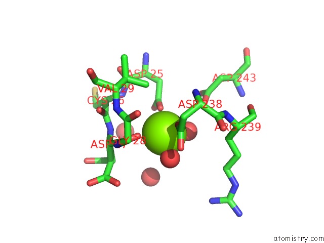
Mono view
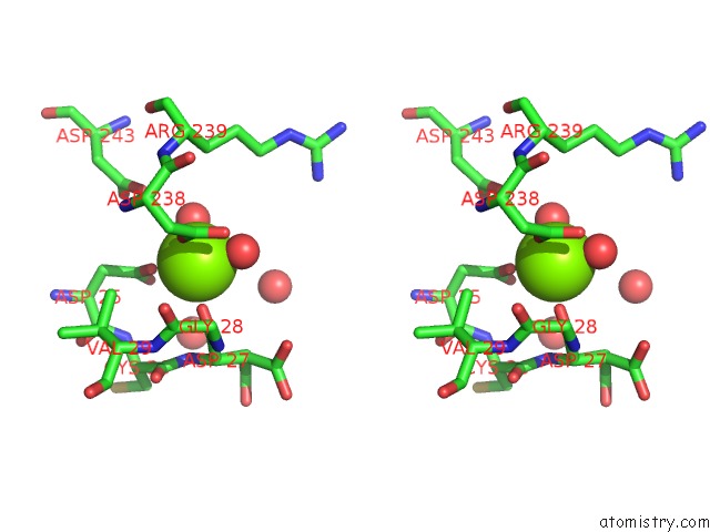
Stereo pair view
Mono view
Stereo pair view
A full contact list of Magnesium with other atoms in the Mg binding
site number 1 of Crystal Structure of Human Pyridoxal 5'-Phosphate Phosphatase within 5.0Å range:
|
Magnesium binding site 2 out of 2 in 2cfs
Go back to
Magnesium binding site 2 out
of 2 in the Crystal Structure of Human Pyridoxal 5'-Phosphate Phosphatase
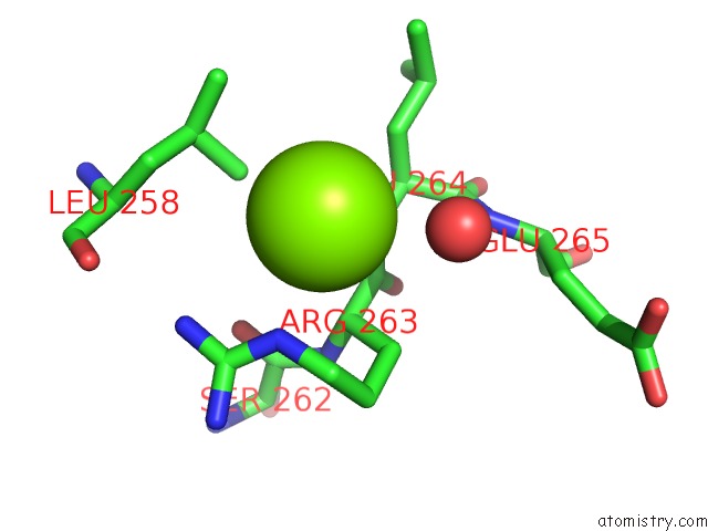
Mono view
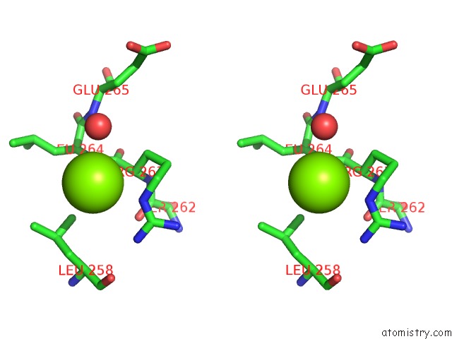
Stereo pair view
Mono view
Stereo pair view
A full contact list of Magnesium with other atoms in the Mg binding
site number 2 of Crystal Structure of Human Pyridoxal 5'-Phosphate Phosphatase within 5.0Å range:
|
Reference:
B.S.Kang,
H.J.Cho,
K.J.Kim,
O.S.Kwon.
Crystal Structure of Human Pyridoxal 5'-Phosphate Phosphatase To Be Published.
Page generated: Sun Aug 10 10:16:43 2025
Last articles
Mg in 2X0QMg in 2X03
Mg in 2WZG
Mg in 2WZD
Mg in 2WZC
Mg in 2WZB
Mg in 2WZ8
Mg in 2WX5
Mg in 2WWR
Mg in 2WW8