Magnesium »
PDB 3a0u-3abk »
3a8m »
Magnesium in PDB 3a8m: Crystal Structure of Nitrile Hydratase Mutant Y72F Complexed with Trimethylacetonitrile
Enzymatic activity of Crystal Structure of Nitrile Hydratase Mutant Y72F Complexed with Trimethylacetonitrile
All present enzymatic activity of Crystal Structure of Nitrile Hydratase Mutant Y72F Complexed with Trimethylacetonitrile:
4.2.1.84;
4.2.1.84;
Protein crystallography data
The structure of Crystal Structure of Nitrile Hydratase Mutant Y72F Complexed with Trimethylacetonitrile, PDB code: 3a8m
was solved by
Y.Yamanaka,
K.Hashimoto,
A.Ohtaki,
K.Noguchi,
M.Yohda,
M.Odaka,
with X-Ray Crystallography technique. A brief refinement statistics is given in the table below:
| Resolution Low / High (Å) | 25.00 / 1.32 |
| Space group | C 1 2 1 |
| Cell size a, b, c (Å), α, β, γ (°) | 114.280, 60.110, 81.811, 90.00, 125.06, 90.00 |
| R / Rfree (%) | 15.7 / 19 |
Other elements in 3a8m:
The structure of Crystal Structure of Nitrile Hydratase Mutant Y72F Complexed with Trimethylacetonitrile also contains other interesting chemical elements:
| Iron | (Fe) | 1 atom |
Magnesium Binding Sites:
The binding sites of Magnesium atom in the Crystal Structure of Nitrile Hydratase Mutant Y72F Complexed with Trimethylacetonitrile
(pdb code 3a8m). This binding sites where shown within
5.0 Angstroms radius around Magnesium atom.
In total 3 binding sites of Magnesium where determined in the Crystal Structure of Nitrile Hydratase Mutant Y72F Complexed with Trimethylacetonitrile, PDB code: 3a8m:
Jump to Magnesium binding site number: 1; 2; 3;
In total 3 binding sites of Magnesium where determined in the Crystal Structure of Nitrile Hydratase Mutant Y72F Complexed with Trimethylacetonitrile, PDB code: 3a8m:
Jump to Magnesium binding site number: 1; 2; 3;
Magnesium binding site 1 out of 3 in 3a8m
Go back to
Magnesium binding site 1 out
of 3 in the Crystal Structure of Nitrile Hydratase Mutant Y72F Complexed with Trimethylacetonitrile

Mono view
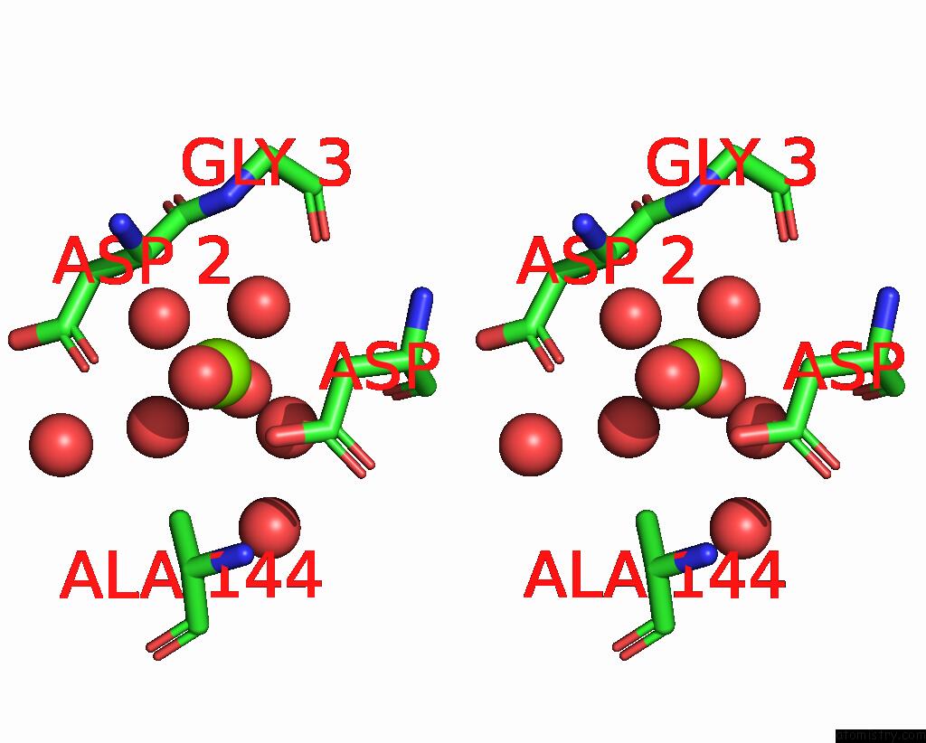
Stereo pair view
Mono view
Stereo pair view
A full contact list of Magnesium with other atoms in the Mg binding
site number 1 of Crystal Structure of Nitrile Hydratase Mutant Y72F Complexed with Trimethylacetonitrile within 5.0Å range:
|
Magnesium binding site 2 out of 3 in 3a8m
Go back to
Magnesium binding site 2 out
of 3 in the Crystal Structure of Nitrile Hydratase Mutant Y72F Complexed with Trimethylacetonitrile

Mono view

Stereo pair view
Mono view
Stereo pair view
A full contact list of Magnesium with other atoms in the Mg binding
site number 2 of Crystal Structure of Nitrile Hydratase Mutant Y72F Complexed with Trimethylacetonitrile within 5.0Å range:
|
Magnesium binding site 3 out of 3 in 3a8m
Go back to
Magnesium binding site 3 out
of 3 in the Crystal Structure of Nitrile Hydratase Mutant Y72F Complexed with Trimethylacetonitrile

Mono view

Stereo pair view
Mono view
Stereo pair view
A full contact list of Magnesium with other atoms in the Mg binding
site number 3 of Crystal Structure of Nitrile Hydratase Mutant Y72F Complexed with Trimethylacetonitrile within 5.0Å range:
|
Reference:
Y.Yamanaka,
K.Hashimoto,
A.Ohtaki,
K.Noguchi,
M.Yohda,
M.Odaka.
Kinetic and Structural Studies on Roles of the Serine Ligand and A Strictly Conserved Tyrosine Residue in Nitrile Hydratase J.Biol.Inorg.Chem. V. 15 655 2010.
ISSN: ISSN 0949-8257
PubMed: 20221653
DOI: 10.1007/S00775-010-0632-3
Page generated: Sun Aug 10 17:23:42 2025
ISSN: ISSN 0949-8257
PubMed: 20221653
DOI: 10.1007/S00775-010-0632-3
Last articles
Mg in 3JAYMg in 3JB3
Mg in 3JB2
Mg in 3JAP
Mg in 3JAM
Mg in 3JAT
Mg in 3JAW
Mg in 3JAS
Mg in 3JAR
Mg in 3J81