Magnesium »
PDB 4to0-4tyq »
4tq9 »
Magnesium in PDB 4tq9: Crystal Structure of A Gdp-Bound G12V Oncogenic Mutant of Human Gtpase Kras
Protein crystallography data
The structure of Crystal Structure of A Gdp-Bound G12V Oncogenic Mutant of Human Gtpase Kras, PDB code: 4tq9
was solved by
J.C.Hunter,
A.Manandhar,
D.Gurbani,
Z.Chen,
K.D.Westover,
with X-Ray Crystallography technique. A brief refinement statistics is given in the table below:
| Resolution Low / High (Å) | 37.05 / 1.49 |
| Space group | C 1 2 1 |
| Cell size a, b, c (Å), α, β, γ (°) | 65.320, 41.427, 115.119, 90.00, 105.09, 90.00 |
| R / Rfree (%) | 16.4 / 19.4 |
Magnesium Binding Sites:
The binding sites of Magnesium atom in the Crystal Structure of A Gdp-Bound G12V Oncogenic Mutant of Human Gtpase Kras
(pdb code 4tq9). This binding sites where shown within
5.0 Angstroms radius around Magnesium atom.
In total 2 binding sites of Magnesium where determined in the Crystal Structure of A Gdp-Bound G12V Oncogenic Mutant of Human Gtpase Kras, PDB code: 4tq9:
Jump to Magnesium binding site number: 1; 2;
In total 2 binding sites of Magnesium where determined in the Crystal Structure of A Gdp-Bound G12V Oncogenic Mutant of Human Gtpase Kras, PDB code: 4tq9:
Jump to Magnesium binding site number: 1; 2;
Magnesium binding site 1 out of 2 in 4tq9
Go back to
Magnesium binding site 1 out
of 2 in the Crystal Structure of A Gdp-Bound G12V Oncogenic Mutant of Human Gtpase Kras
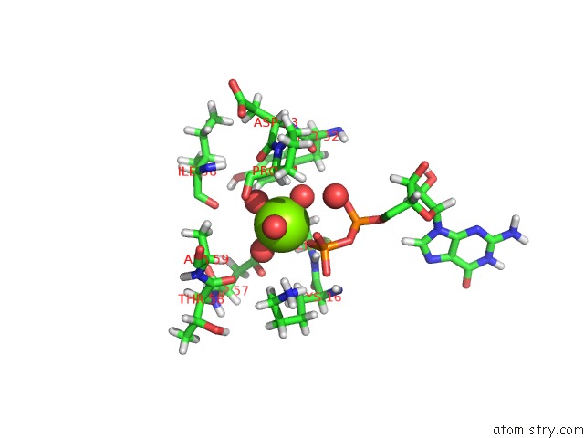
Mono view
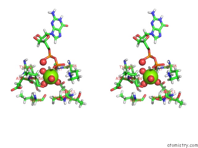
Stereo pair view
Mono view
Stereo pair view
A full contact list of Magnesium with other atoms in the Mg binding
site number 1 of Crystal Structure of A Gdp-Bound G12V Oncogenic Mutant of Human Gtpase Kras within 5.0Å range:
|
Magnesium binding site 2 out of 2 in 4tq9
Go back to
Magnesium binding site 2 out
of 2 in the Crystal Structure of A Gdp-Bound G12V Oncogenic Mutant of Human Gtpase Kras
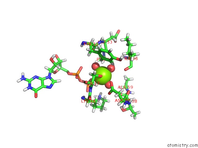
Mono view
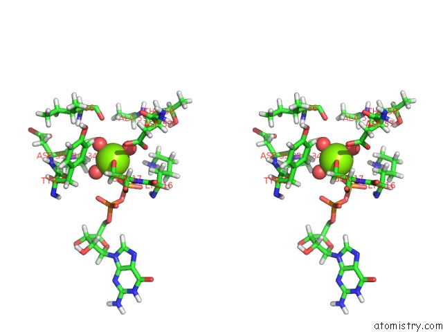
Stereo pair view
Mono view
Stereo pair view
A full contact list of Magnesium with other atoms in the Mg binding
site number 2 of Crystal Structure of A Gdp-Bound G12V Oncogenic Mutant of Human Gtpase Kras within 5.0Å range:
|
Reference:
J.C.Hunter,
A.Manandhar,
M.A.Carrasco,
D.Gurbani,
S.Gondi,
K.D.Westover.
Biochemical and Structural Analysis of Common Cancer-Associated Kras Mutations. Mol Cancer Res. V. 13 1325 2015.
ISSN: ESSN 1557-3125
PubMed: 26037647
DOI: 10.1158/1541-7786.MCR-15-0203
Page generated: Tue Aug 12 00:10:02 2025
ISSN: ESSN 1557-3125
PubMed: 26037647
DOI: 10.1158/1541-7786.MCR-15-0203
Last articles
Mg in 5AIVMg in 5AIS
Mg in 5AHN
Mg in 5AHK
Mg in 5AFX
Mg in 5AEL
Mg in 5AES
Mg in 5AGA
Mg in 5AC1
Mg in 5ADU