Magnesium »
PDB 6j7r-6jlj »
6jd4 »
Magnesium in PDB 6jd4: Atpase
Protein crystallography data
The structure of Atpase, PDB code: 6jd4
was solved by
S.H.Wang,
J.Li,
Z.H.Rao,
with X-Ray Crystallography technique. A brief refinement statistics is given in the table below:
| Resolution Low / High (Å) | 43.72 / 2.10 |
| Space group | P 21 21 21 |
| Cell size a, b, c (Å), α, β, γ (°) | 61.220, 77.314, 106.011, 90.00, 90.00, 90.00 |
| R / Rfree (%) | 20.2 / 24.6 |
Magnesium Binding Sites:
The binding sites of Magnesium atom in the Atpase
(pdb code 6jd4). This binding sites where shown within
5.0 Angstroms radius around Magnesium atom.
In total 2 binding sites of Magnesium where determined in the Atpase, PDB code: 6jd4:
Jump to Magnesium binding site number: 1; 2;
In total 2 binding sites of Magnesium where determined in the Atpase, PDB code: 6jd4:
Jump to Magnesium binding site number: 1; 2;
Magnesium binding site 1 out of 2 in 6jd4
Go back to
Magnesium binding site 1 out
of 2 in the Atpase
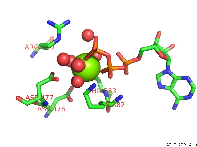
Mono view
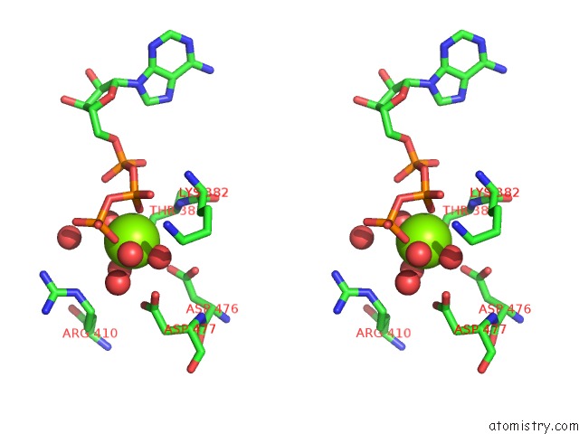
Stereo pair view
Mono view
Stereo pair view
A full contact list of Magnesium with other atoms in the Mg binding
site number 1 of Atpase within 5.0Å range:
|
Magnesium binding site 2 out of 2 in 6jd4
Go back to
Magnesium binding site 2 out
of 2 in the Atpase
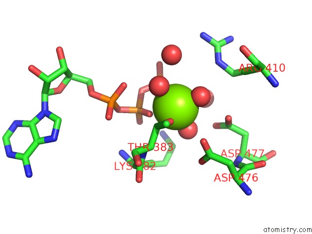
Mono view
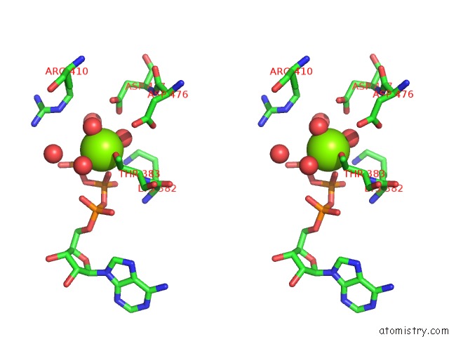
Stereo pair view
Mono view
Stereo pair view
A full contact list of Magnesium with other atoms in the Mg binding
site number 2 of Atpase within 5.0Å range:
|
Reference:
S.Wang,
K.Zhou,
X.Yang,
B.Zhang,
Y.Zhao,
Y.Xiao,
X.Yang,
H.Yang,
L.W.Guddat,
J.Li,
Z.Rao.
Structural Insights Into Substrate Recognition By the Type VII Secretion System. Protein Cell 2019.
ISSN: ESSN 1674-8018
PubMed: 31758528
DOI: 10.1007/S13238-019-00671-Z
Page generated: Wed Aug 13 09:10:09 2025
ISSN: ESSN 1674-8018
PubMed: 31758528
DOI: 10.1007/S13238-019-00671-Z
Last articles
Mg in 7A17Mg in 7A0V
Mg in 6ZZX
Mg in 6ZXS
Mg in 7A0Q
Mg in 7A0P
Mg in 7A0C
Mg in 721P
Mg in 6ZXH
Mg in 6ZXG